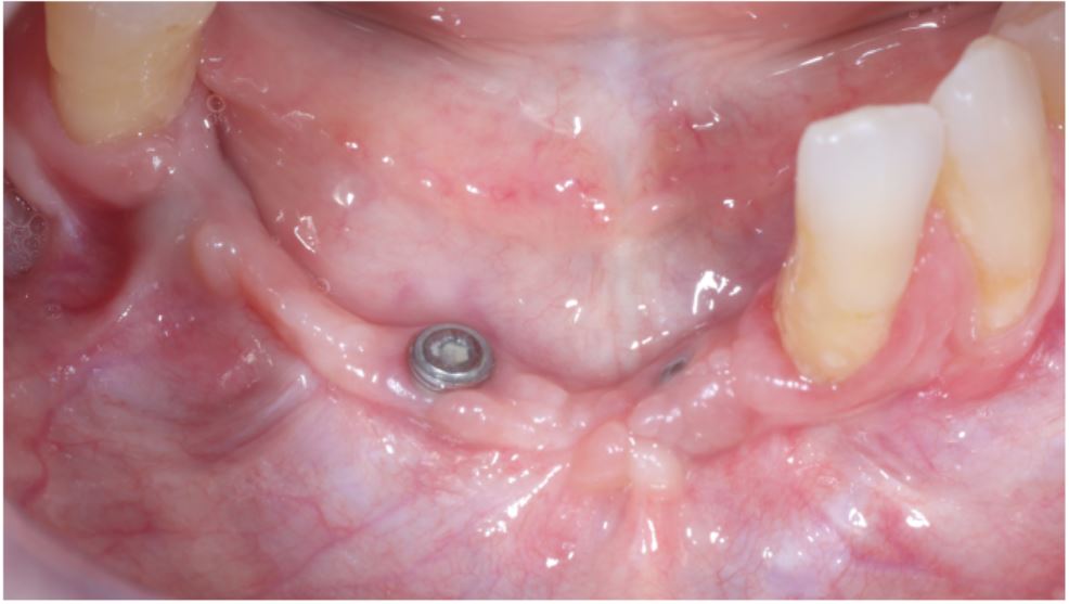
Figure 1: Initial picture showing absence of keratinized tissue around three submerged implants on anterior mandible that will be prosthetically rehabilitaded
Figure 1: Initial picture showing absence of keratinized tissue around three submerged implants on anterior mandible that will be prosthetically rehabilitaded
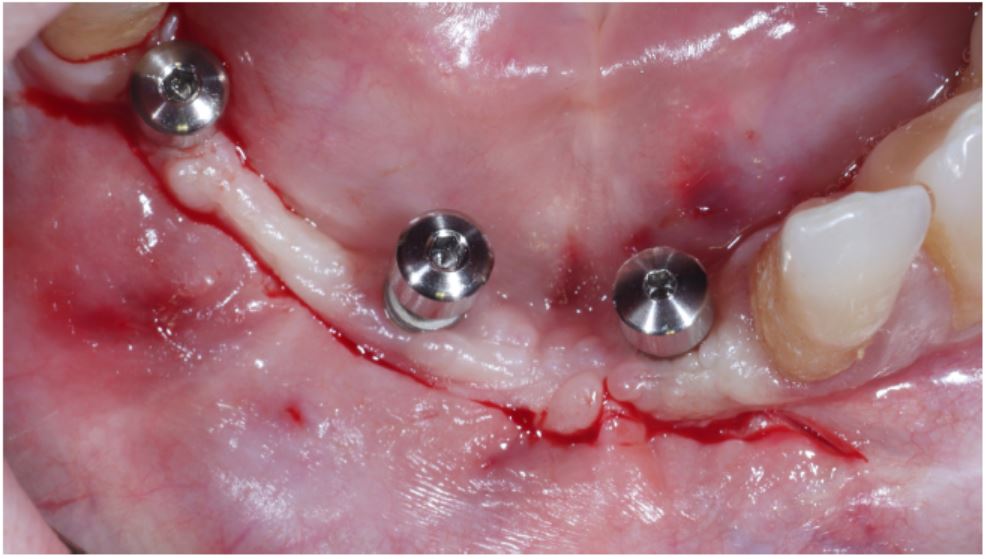
Figure 2: Initial incision for flap elevation delimiting the region that will receive the graft
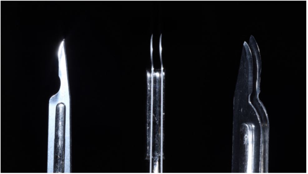
Figure 3: Division of partial thickness flap unhanding intact periosteum attached to bone
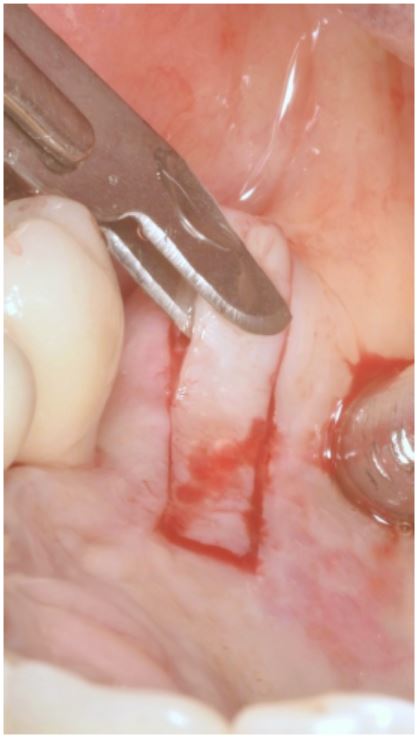
Figure 4: Double blade scapel mounted. Measurement of recipient area was done previously using a calibrated periodontal probe. A template was made with sterilized paper and transferred to the donor site on the palate. A superficial incision surrounding the map is made to outline the graft. A double-blade scalp is used to initate the incision from the mesio-disto distance of the graft
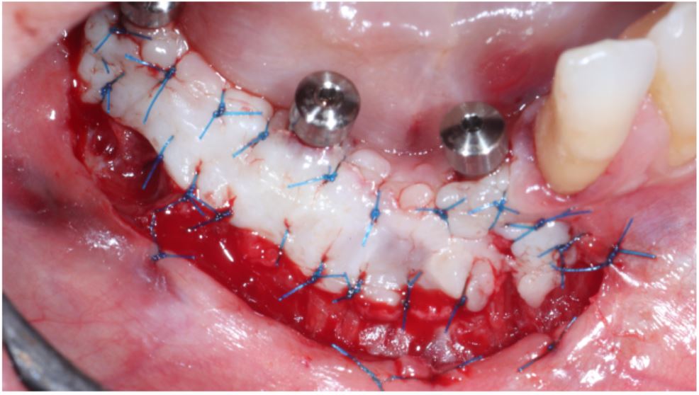
Figure 5: Double blade scapel in position at donor site
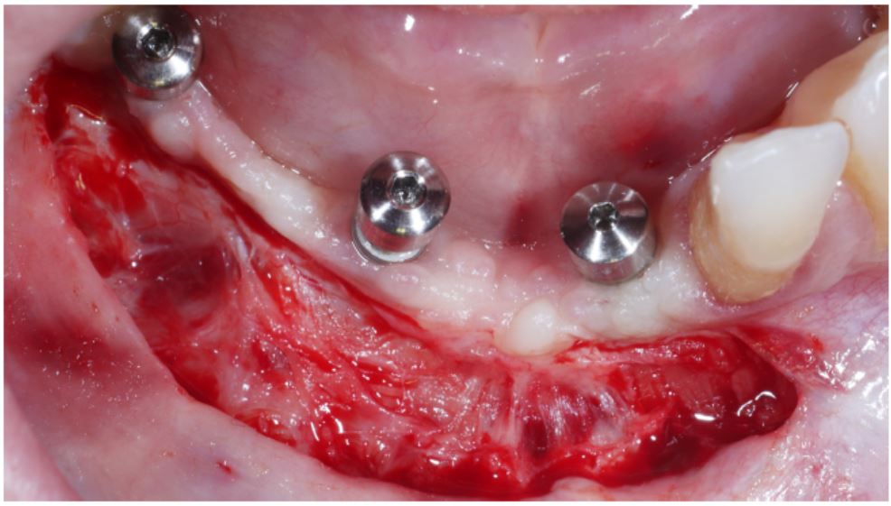
Figure 6: Double blade scapel in position removing gingival graft
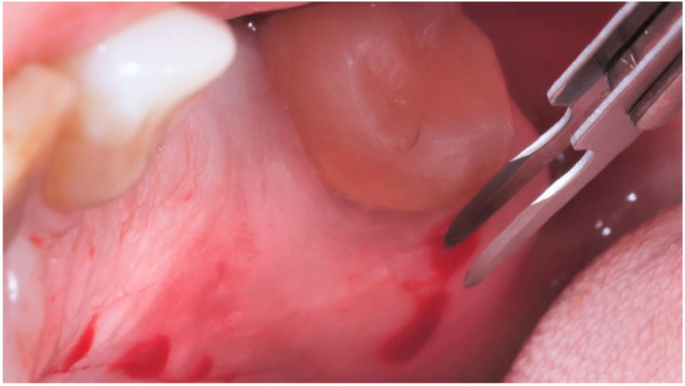
Figure 7: Donor site after graft removal and sutured
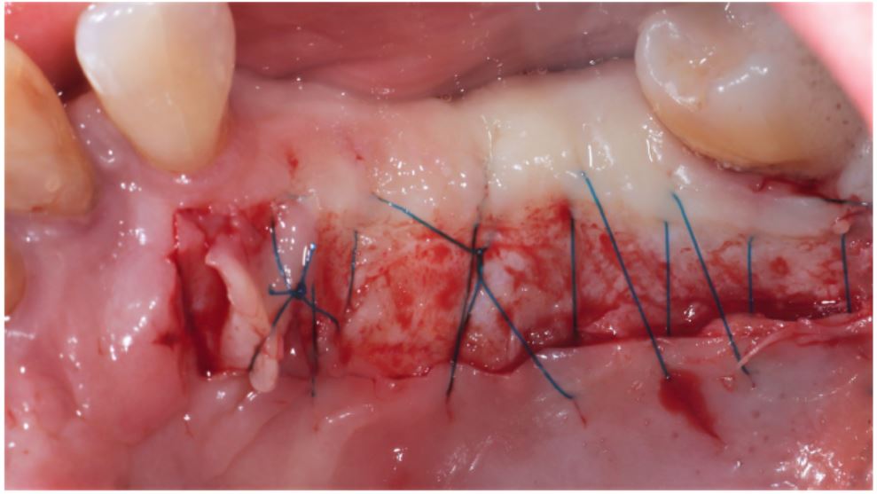
Figure 8: Graft thickness checked
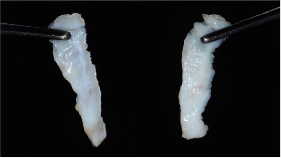
Figure 9: Free gingival graft in position to check measurements
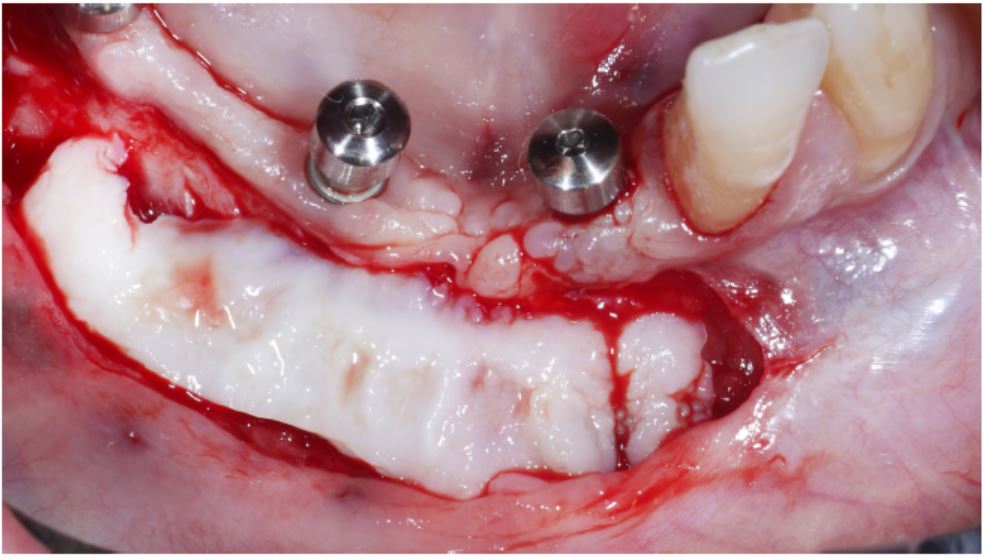
Figure 10: Graft sutured at position
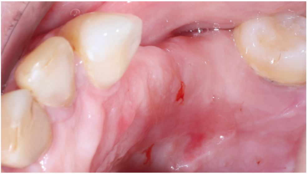
Figure 11: Donor site with 30 days healing
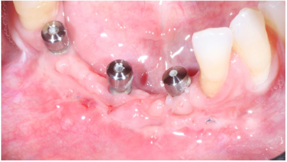
Figure 12: Receptor area after 30 days healing
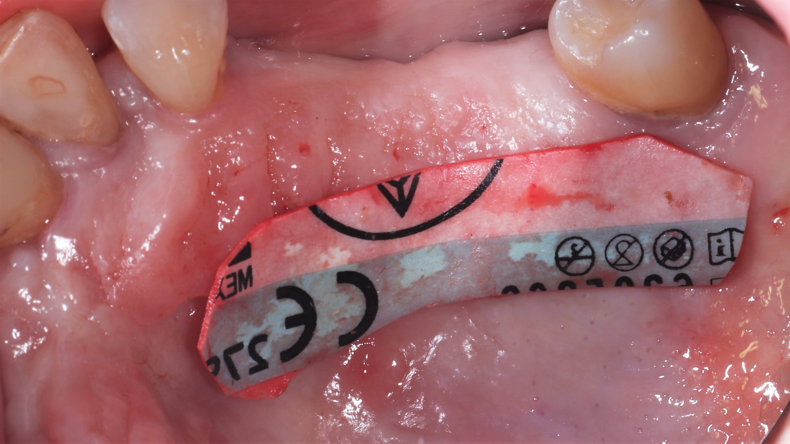
Figure 13: Donor Site
Tables at a glance
Figures at a glance