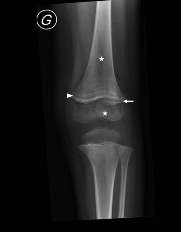
Figure 1 Left knee radiograph demonstrating multiple clear metaphyseal bands in the distal femur (white arrow head) associated with irregular and enlarged metaphyseal margin (white arrow). Diffuse osteopenia (stars) is also noted.
Figure 1 Left knee radiograph demonstrating multiple clear metaphyseal bands in the distal femur (white arrow head) associated with irregular and enlarged metaphyseal margin (white arrow). Diffuse osteopenia (stars) is also noted.
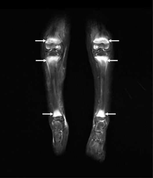
Figure 2 Bilateral lower leg MRI: coronal fat-suppressed T2-weighted image revealing bilateral intra-osseous edematous changes of the metaphyseal area (white arrows) of the ankles and knees. Edematous changes were also noted in the distal epiphysis of the femur bilaterally (stars). Whole-body MRI of this patient shows multiple similar anomalies in the wrists and shoulders.
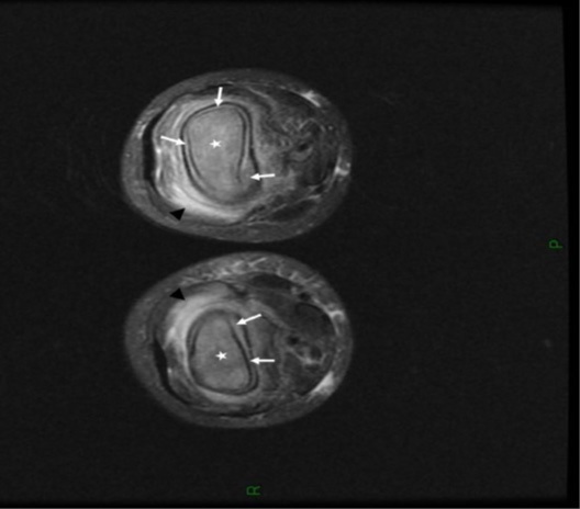
Figure 3Bilateral knee MRI: axial fat-suppressed T2-weighted image showing important bilateral edematous changes within (stars) and around the bone (black arrowheads) associated with bilateral circumferential subperiosteal collections (white arrows) related to bilateral subperiosteal hematomas.