Hematology as an Analytical Tool in Forensic Science
Received Date: August 3, 2021 Accepted Date: August 5, 2021 Published Date: September 7, 2021
doi: 10.17303/jfrcs.2021.6.103
Citation: Gupta K (2021) Hematology as an Analytical Tool in Forensic Science. J Forensic Res Crime Stud 6: 1-8.
Abstract
Blood is one of the most important biological evidence found at a crime scene. In a violent crime, the blood sample obtained may belong to the victim, the suspect(murderer) or any other eyewitness. A proper analysis of blood samples obtained at a crime scene may contribute to clarify the circumstances under which the crime would have been committed. The information obtained by studying the blood sample may point the criminal investigation in the right direction and can help solve the crime. Hematology refers to the study of blood and it can play an important tool in analyzing the crime. The blood analysis begins with the presumptive tests to confirm that a given sample is blood. Then precipitin technology may be used to find the species to which the blood belongs. Methods like DNA fingerprinting may be used to match the blood at crime scene with the blood of suspect to get a step closer to finding the criminal. We report here few of the analysis of blood that can be done to possibly fasten the process of case solving and find the right criminal.
Keywords: Forensic Science; Blood Group Typing; Blood Detection; Precipitin Technology; Blood Spatter Analysis; Mosquito Blood Analysis
Highlights- Bioidentification
- Visualization technique to confirm the presence of blood
- Precipitin technology to find if blood belongs to Human or not.
Introduction
Forensic serology is the study and individualization of various body fluids (blood,semen,saliva,urine, vomit,fecal matter) found at the crime scene and establishing its relationship with the crime scene to proceed the case in the right direction [1]. Blood, a major part of the complex transporting system of the body, is composed of corpuscular elements (erythrocytes, leukocytes and thrombocytes) suspended in light yellow coloured liquid called plasma.
Blood forms around 8% of the total body weight. Plasma forms 55% of the total volume of blood and is composed of 90% water and other biochemicals such as proteins, nutrients and other waste products formed during metabolism.
The corpuscular elements or formed elements in blood include erythrocytes, leukocytes and thrombocytes. Erythrocytes are tiny (7.8 microns diameter) non - nucleated, biconcave shaped cells. Erythropoietin controls the production of erythrocytes in the body. The principle function of Red blood cells is oxygen transport by binding the oxygen to hemoglobin molecules. Leukocytes are cells larger than erythrocytes but fewer in number in the body. The average diameter of leukocytes is 12-17 microns. They play a major role in generating immune response.
Thrombocytes are small a nucleated, disk shaped fragments of large cells called megakaryocytes and have a diameter of around 2-4 microns are responsible for clotting response [2] (Table 1).
The erythrocytes have some special proteins called antigens on their surface. These antigens are responsible for the phenomenon of blood typing. There are approximately 100 different types of antigens found on the surface of erythrocytes including the rare antigens. Out of these 100 antigens, there are around 30 commonly occurring antigens.
The scientists discovered more than 100 antigens and around 23 blood types by 1937 [3]. By 2014, the international society of blood transfusion could recognize 33 different blood types [4].
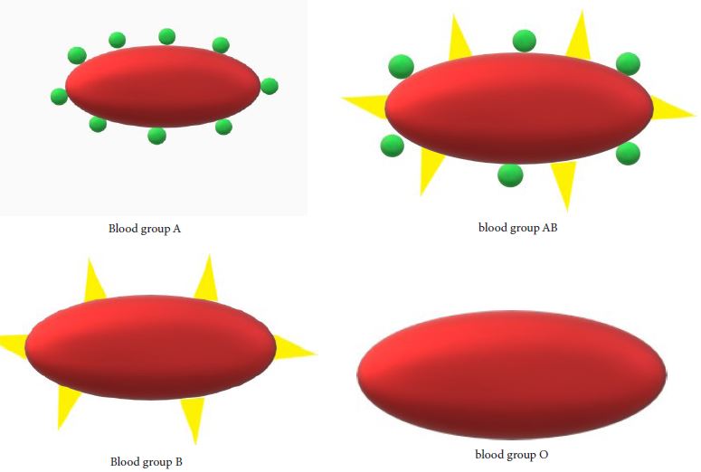
The commonly used blood group system is the ABO blood group system which was discovered by Landsteiner in 1900. The ABO system includes A antigens, B antigens and antibodies against them. The ABO blood grouping system thus gives rise to four major blood groups (A,B,AB and O) along with some subgroups which differs in patterns and degree of agglutination [5].
The A antigen has subtype A1 and A2 which further produces two different subgroups of A and AB blood groups. 80% of the individuals with A antigen have A1 subtype and the rest 20% have A2 subtype. The individuals with A1 antigen have A1 or A1B as the blood group and individuals with A2 antigen have A2 or A2B as the blood group. The difference between A1 and A2 antigen is in the reactivity of lectin which means The Anti A1 present as a cold agglutinin and agglutinates the A1 cells. The anti A1 is possessed by around 0.4% of A2 and 25% of A2B subgroups [6].
The blood type needs to be checked and only compatible blood types can be transfused in blood transfusion. Transfusion of incompatible blood type can prove to be fatal or may cause complications in the body of the receiver. This is due to the immunological effect of A and B antigens and the production of the corresponding antibody in their presence [7] (Table 2).
BIO identification
The blood type can be used to determine the paternity of children and also to prove innocence of a person. Due to large numbers of people possessing the same blood type (ABO BLOOD TYPING), the blood type of the blood sample obtained at a crime scene cannot be individualized. A suspect might have the same blood type as that of the blood obtained at a crime scene, but this cannot be used to prove guilt. Nevertheless blood type identification can be used to establish innocence. The people from the suspected population with a different blood type can be excluded from suspicion as the source of the blood sample at the crime scene. Blood group testing was used by forensic scientists uptil 1980s, after which DNA testing was performed because of its accuracy and reliability [8] (Table 3).
Presumptive Tests for Blood Identification
The wet blood found at the crime scene can easily be identified as blood just by looking at it. In case an old, dried blood is found at the crime scene, it may not be easy enough to find whether it’s blood or not. Also, if the bloodstain is not visible or if it is found on a dark material, then the presumptive tests need to be conducted in order to assure whether the sample found is a blood sample or is it something else. The chemical methods to detect blood can be divided into 3 categories:
1.Crystal test - this test is based on the ability of haem molecules to react with certain reagents like pyridine to form crystals.
2.Catalytic test - this test is based on the catalytic property of haem molecules to breakdown hydrogen peroxide.
3.Instrumental methods - In these methods, chromatography is used to detect presence of haemoglobin to identify blood [9].
Crystal tests
The basis of this test is that the haem molecules react with certain reagents to produce haemoglobin derivative crystals such as haematin, haemin and haemochromogen.The crystal formation is observed microscopically. Teichmann was the first to produce haemin crystals in 1853. Since then, the crystal tests were widely used for detection of blood. But the test sometimes is not successful due to overheating, the impurities in acid, inhibitory effect of rust, fat etc. or change in the blood composition. This led to the introduction of various other modifications in the Teichmann method. In 1909, Donogany and Burker found that pyridine could be used to produce haemochromogen crystals to detect blood [10].
Pyridine alone gave false positive results. Thus in 1912, Takayama suggested a solution containing pyridine which gave the correct results. The composition of this solution is pyridine, 10% sodium hydrate solution, saturated solution of dextrose and distilled water. Pyridine forms pink to salmon pink colored crystals of Pyridine ferroprotoporphyrin in the presence of haem molecules, when heated. The shape of the crystals varies due to the freshness of blood and time of formation. The shape may be flat, rhomboid or curved and sometimes there is a sheaf formation [11]. The pyridine forms the Haemochromogen crystals by binding to the haem molecules. Oxygen competes with pyridine for the same binding site. So to increase the rate of formation of haemochromogen crystals, the oxygen scavenger such as dithiothreitol (Cleland’s reagent) is used [12].
Takayama test

Nitrogenous bases including methylamine, nicotine, histidine and glycine can also be used as a reagent in crystal tests. Bloodstains upto 20 years old can give a positive crystal test.
Catalytic tests
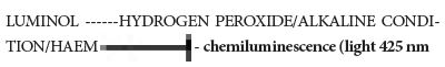
Catalytic tests are based on the catalytic, peroxidase - like activity of the heme group of haemoglobin. The breakdown of hydrogen peroxide produces an oxidizing species which further oxidizes a substrate to produce a color change. The commonly used substrates are benzidine, ortho - tolidine , leucomalachite green, leucocrystal violet and phenolphthalein. Alternatively, 3-aminophthalhydrazide (luminol) is made to react with haemoglobin and hydrogen peroxide to produce a product that luminesces [13].
The Kastle meyer test is widely used to detect blood. The Kastle Meyer test requires two solutions for detection of blood. First is the solution of hydrogen peroxide of twenty volumes. The second solution is composed of phenolphthalein, potassium hydrate and distilled water. In the kastle meyer test, a colourless solution of reduced phenolphthalein (in aqueous solution) and zinc is made to react with the sample and hydrogen peroxide. An immediate colour change to a bright pink colour indicates the presence of blood in the sample [14].
Reduced phenolphthalein (colorless) + haemoglobin 3% HYDROGEN

Luminol test is based on the production of a chemiluminescence when luminol is oxidized by haemoglobin. This technique is sensitive to blood stains and does not give a positive result with other biological fluids. Luminol test is majorly used to detect the blood stains that have been tried to be erased by the offender. But it cannot be used to detect blood stains on dark fabric as this could lead to DNA degradation in the blood sample. One thing that needs to be kept in mind is that since luminol gives chemiluminescence in the presence of blood. Thus it can only be used in a dark room because only then the luminescence would be visible [15].
One such commonly used test is the sangur test. The sangur sticks have the reagent in an immobilized form. When the sangur sticks are rubbed gently on the stain and moistened, a color change from pale yellow to intense greenish blue indicates the presence of blood [9].
Luminol test
The major limitation of the catalytic tests is that since they are based on the peroxidase like activity. So a false positive result can be obtained in the presence of catalase or Peroxidase (present in plant and animal material), oxidizing chemicals and metals (copper and iron). Combining a catalytic test with any other test can be used to overcome the possibility of false positive results.
Instrumental methods
High performance liquid chromatography is used for detecting the presence of blood in a sample using the absorbance of haemoglobin. Colorimetric method is performed to determine the relative percentage of haemoglobin in a given sample. This helps to find out whether sufficient haemoglobin is present in a sample that it could be a blood sample or not [16]. Determination of Haemoglobin can be done by first separating haemoglobin on cellulose acetate electrophoresis pH 8.9, microcolumn chromatography followed by analysis of elution with spectrometry and high performance liquid chromatography (HPLC) HPLC (high performance liquid chromatography). HPLC is a sensitive method for identifying HbA2 (adult haemoglobin) or HbF (fetal haemoglobin). Presence of Haemoglobin indicates the presence of blood [17].
Precipitin technology
Once a sample found at a crime scene is labelled as a blood sample, the next important aspect to understand is the species to which the blood sample belongs. This biological differentiation between the blood samples from different species of animals can only be known by a test known as precipitin test [18]. Fresh blood samples can be easily differentiated and it can be identified if the blood is mammalian, reptilian or avian blood. The task of differentiating the mammalian blood between different species can only be fulfilled by performing precipitin test.
The precipitin test is based on the formation of a precipitum when a diluted serum reacts with its antiserum. To test whether a blood sample is a human blood or not, anti human serum is used.
To perform the qualitative method of the precipitin test firstly the dried blood was immersed in a small quantity of distilled water for a few hours so that an extract is obtained. Afterwards, 1.2% salt solution was added to the extract.
Small quantities of the extract were taken in a few test tubes with a black background. Gradually anti human serum and some other anti serum was added in different test tubes containing the blood sample. Antiserum being a solution with a greater specific gravity flows at the bottom of the test tube. The test tube in which a corresponding antiserum was added developed a white cloud at the junction of the two fluids which gradually formed a precipitum at the bottom of the test tube. The limitation with the qualitative test is that if the antiserum is powerful then it may precipitate out in the presence of serum of the distantly related animals also.
In forensic science, oftenly the qualitative method is performed to check if the blood obtained is human blood or belongs to some other animal [19].
Mosquito blood to find suspect
Sometimes mosquitoes can be obtained at the crime scene. A detailed study of the last meal of a mosquito can be really useful in determining the time at which the mosquito bit the person by studying the degree of human DNA digestion and also help to determine the source of human DNA in the mosquito. This technique has often helped the forensic scientists to catch the criminal. It is been observed that in many mosquitoes species like Culex pipiens pallens and Aedes albopictus, the genotyping can be done until 2 days of post feeding as the blood meal takes about 3 days to get completely digested. Some forensic scientists use the precipitin technology for the specie identification of the blood meal. Classic ABO blood typing and serum typing meth ods are used for group-specific component (Gc) and haptoglobin (Hp), and red cell enzyme typing for phosphoglucomutase (PGM), acid phosphatase (AcP), and esterase D. Identifying the host species using the cytochrome b and/or cytochrome oxidase genes in the mitochondrial DNA (mtDNA) by direct sequencing and polymerase chain reaction with the restriction fragment length polymorphisms (PCR-RFLPs) are some of the DNA based techniques for determining host species. Once the host specie is identified, the human DNA found from the blood meal can be individualized using variable number tandem repeats or short tandem repeats genotyping using gel electrophoresis followed by silver staining. Nowadays multiplex STR typing kits are usually used for human DNA individualization [20].
Blood Spatter Analysis
In a violent crime, often the blood spatters are found on the crime scene. The analysis of the blood spatters can help the forensic scientists to reconstruct the crime scene and make the case go in a positive direction.
Blood spatter analysis can have major drawbacks if a large number of blood stains are found at the crime scene. But sometimes blood spatter analysis can help the serologists to individualize the blood stains found at the crime scene by deeply studying the blood spatter pattern [21]. Earlier the trigonometric calculations of the individual blood stain was done manually to understand the blood stain pattern. Advancements in science have led to the introduction of a 3D scanning method that makes the study of blood spatter analysis easier and error less [22]. The blood spatter analysis is done by examining the shape, location and distribution of blood stain patterns. In a blood spatter analysis, few things that are analysed are distance of the blood source from the target, impact angles, weapon used to shed blood, nature of force used to cause blood shed and interpreting the transfer patterns. Usually the greater the force applied to cause blood shed, the smaller would be the spatter [23].
Conclusion
A wet blood sample found at the crime scene may be regarded as blood even by looking but a dried blood sample may not be easily designated as blood. Various presumptive tests like crystal test, instrumental methods and catalytic tests may be used to detect blood. Precipitin technology is used to analyse the species of organism to which the blood belongs. The blood sample found at a crime scene may not always belong to a human. It can belong to any species from the Animalia kingdom. Sometimes the mosquito found at the crime scene may reveal the suspect if it has bit the suspect. The blood from the stomach of the mosquito can be analysed to find any possible clue. The blood spatter analysis is done to recreate the crime scene and find the possible ways in which the crime could have been committed
Acknowledgement
I would like to express my deep gratitude for the college principal, Dr. Savita Roy for her support.
- Pokupcic K (2017) Blood as an important tool in crime investigation. Journal of Forensic Sciences and Criminal Investigation 10.19080/JFSCI.2017.03.555615.
- Chargé S, Hodgkinson K (2017) Blood: The basics Online publication date, India.
- Corey H (2016) ABO Blood Type Identification and Forensic Science (1900-1960), Embryo Project Encyclopedia, USA.
- Mitra R, Mishra N, Rath GP (2014) Blood groups systems. Indian J Anaesth 58: 524-8.
- Yamamoto F (2004) Review: ABO blood group system--ABH oligosaccharide antigens, anti-A and anti-B, A and B glycosyltransferases, and ABO genes. Immunohematology 20: 3-22.
- Giriyan SS, Agrawal A, Bajpai R, Nirala NK (2017) A1 and A2 Sub-Types of Blood Group ‘A’: A Reflection of their Prevalence in North Karnataka Region. J Clin Diagn Res 11: EC40-EC42.
- Dean L (2017) ABO Blood Group In: Pratt VM, Scott SA, Pirmohamed M, Esquivel B, Kane MS, Kattman BL, Malheiro AJ, editors. Medical Genetics Summaries [Internet]. Bethesda (MD): National Center for Biotechnology Information (US), USA.
- Corey H (2016) ABO Blood Type Identification and Forensic Science (1900-1960), Embryo Project Encyclopedia, USA.
- Winchester RV, Wansbrough H (2010) Blood Detection by Chemical Methods, USA.
- Kerr DJ, Mason VH (1926) The Haemochromogen Crystal Test for Blood. Br Med J 1: 134-6.
- Greaves AV (1932) The Use of Takayama’s Solution in the Identification of Blood Stains. Br Med J 1: 932-3.
- Hatch AL (1993) A modified reagent for the confirmation of blood. J Forensic Sci 38: 1502-6.
- National Forensic Science Technology Center (2011) Identification and grouping of bloodstain, USA.
- Glaister J (1926) The Kastle-Meyer Test for the Detection of Blood: Considered from the Medico-Legal Aspect. Br Med J 1: 650-2.
- Vandewoestyne M, Lepez T, Van Hoofstat D, Deforce D (2015) Evaluation of a Visualization Assay for Blood on Forensic Evidence. J Forensic Sci 60: 707-11.
- Haldane J (1901) The colorimetric determination of haemoglobin. J Physiol 26: 497-504.
- George E, Jamal AR, Khalid F, Osman KA (2001) High performance liquid chromatography (HPLC) as a screening tool for classical Beta-thalassaemia trait in malaysia. Malays J Med Sci 8: 40-6.
- Hektoen L (1918) The Precipitin Test For Blood. JAMA 70: 1273-8.
- Graham-Smith G, Sanger F (1903) The Biological or Precipitin Test for Blood considered mainly from its medico-legal aspect. Journal of Hygiene 3: 258-91.
- Hiroshige Y, Hara M, Nagai A, Hikitsuchi T, Umeda M, et al. (2017) A human genotyping trial to estimate the post-feeding time from mosquito blood meals. PLoS ONE 12: e0179319.
- Karger B, Rand S, Fracasso T, Pfeiffer H (2008) Institute of Legal Medicine, University of Münster, Röntgenstrasse 62, 48149 Münster, Germany Bloodstain pattern analysis—Casework experience 10.1016/j.forsciint.2008.07.010.
- Esaias O, Noonan GW, Everist S, Roberts M, Thompson C, et al. (2019) Improved Area of Origin Estimation for Bloodstain Pattern Analysis Using 3D Scanning. J Foren Sci 65: 722-8.
- Crime Scene Forensics, LLC, Concord, NC, USA.
FIGURE 1
Tables at a glance
Figures at a glance