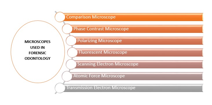
Figure 1: Various Microscopes used in Forensic Odontology
Figure 1: Various Microscopes used in Forensic Odontology
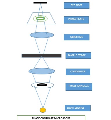
Figure2: Phase Contrast Microscope- Schematic Diagram
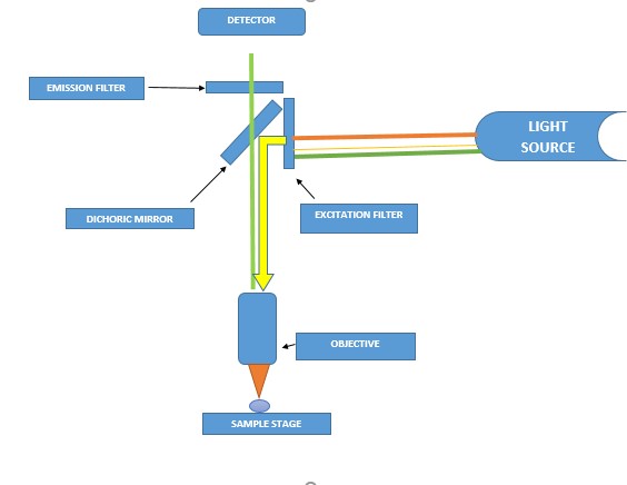
Figure3: Fluorescent Microscope- Schematic Diagram
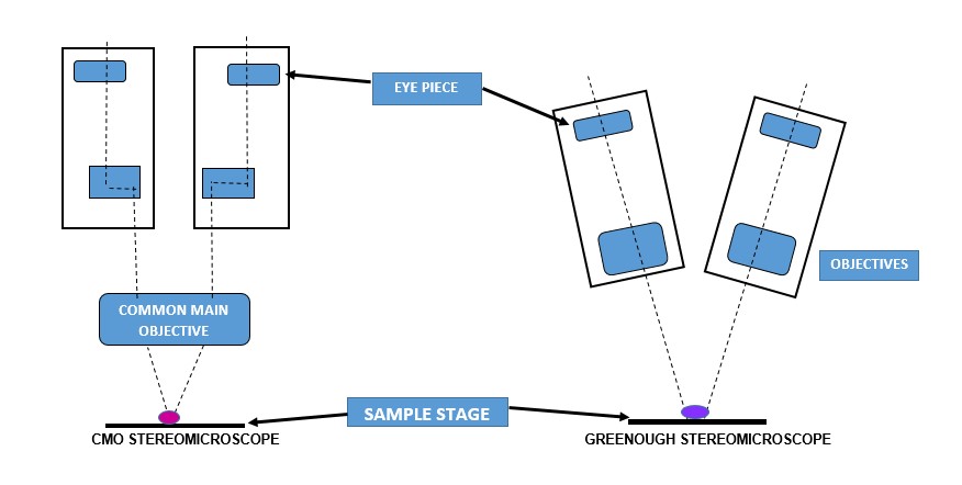
Figure 4: Stereomicroscopes- Types and Schematic Diagram
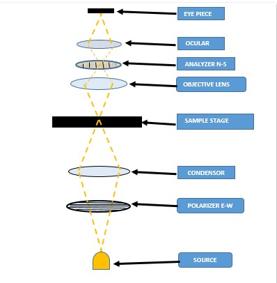
Figure 5: Polarizing Microscope- Schematic Diagram
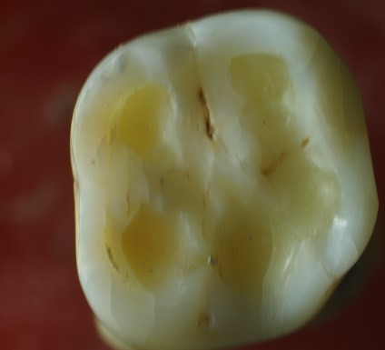
Figure 6: Stereomicroscopic image of Molar tooth showing attrition facets on occlusal surface. (Courtesy- Dr. Harita Paghadal, PG student, GDCHA)
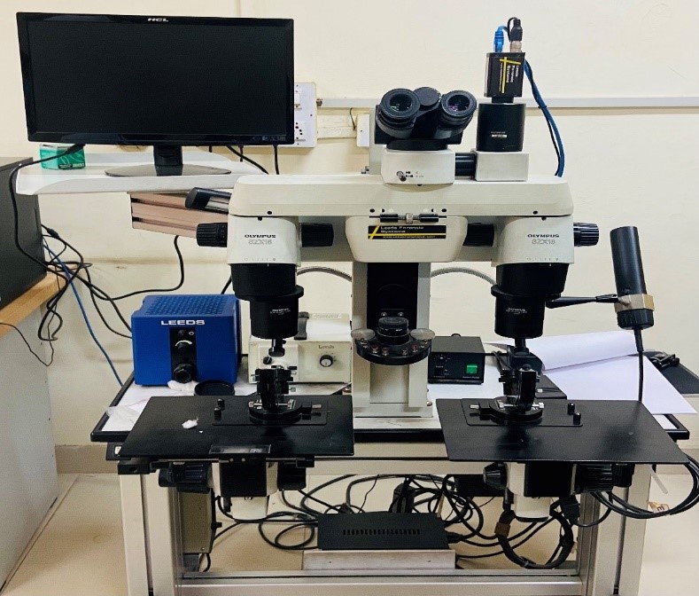
Figure 7: Comparison Microscope
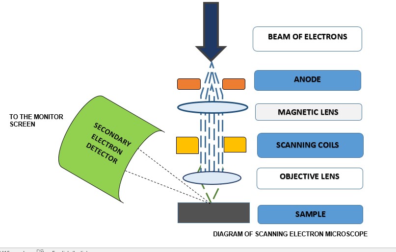
Figure 8: Scanning Electron Microscope- Schematic Diagram
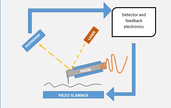
Figure 9: Atomic Force Microscope- Schematic Diagram
Tables at a glance
Figures at a glance