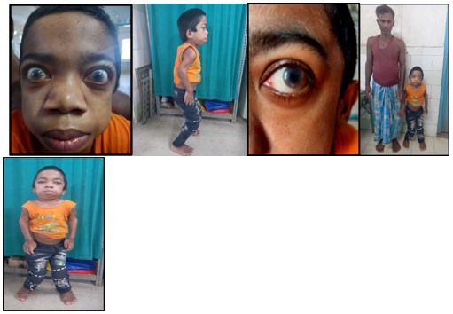
Figure 1: Clinical images of the patient
Figure 1: Clinical images of the patient
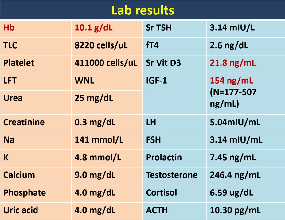
Figure 2: Laboratory investigations
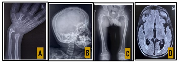
Figure 3: Radiology images
A: X-ray B/L wrist joint: Bone Age = 12-13 yr, suggesting Rickets
B: X-ray Skull (Lat view): Frontal bossing & J shaped sella
C: X-ray of pelvis: Hip dysplasia and femoral epiphyseal widening
D: MRI: Dilated ventricles, Hyperintensities in periventricular, fronto-parietal & temporal white matter.
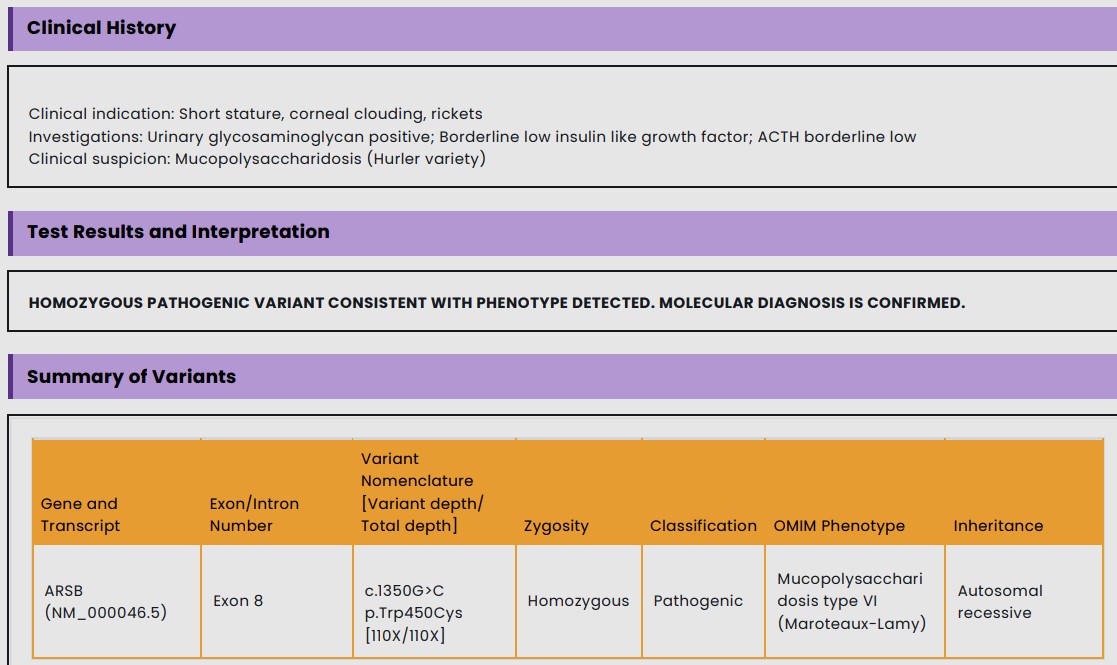
Figure 4: Genetic study
Figure 5
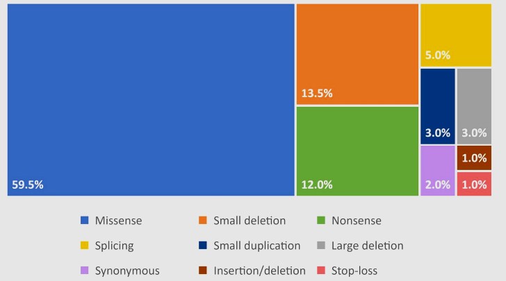
Figure 6: Distribution of Variant Types in the ARSB Gene.
Figures at a glance