Phytochemical-Assisted Synthesis of Nanoparticles from Pongamia pinnata Pods and Their Role in Polymer Composites
Received Date: April 15, 2026 Accepted Date: May 11, 2026 Published Date: May 18, 2026
doi:10.17303/jmsa.2026.10.102
Citation: Praveen Meena, Arun Sharma, Dinesh Kulhary (2026) Phytochemical-Assisted Synthesis of Nanoparticles from Pongamia pinnata Pods and Their Role in Polymer Composites. J Mater sci Appl 10: 1-12
Abstract
Pongamia pinnata pods, a plentiful source of bioactive phytochemicals, have surfaced as a sustainable resource for the eco-friendly synthesis of nanoparticles. This study reports the ecofriendly synthesis of zinc oxide (ZnO) nanoparticles utilizing Pongamia pinnata pod extract and their incorporation into polyvinyl alcohol (PVA) to fabricate functional nanocomposite films. The natural reducing and stabilizing agents provided by the phytochemicals such polyphenols, tannins, and flavonoids in the extract made it a safer option than traditional procedures without sacrificing effectiveness. With clear peaks at 2θ values of 31.8°, 34.5°, and 36.3°, X-ray diffraction (XRD) verified the creation of hexagonal wurtzite ZnO. The average crystallite size was estimated to be 18–25 nm using the Debye–Scherrer equation. Scanning Electron Microscopy (SEM) revealed spherical nanoparticles with diameters ranging from 20–40 nm and moderate agglomeration, likely influenced by phytochemical surface capping. These ZnO nanoparticles were uniformly embedded into a PVA matrix through solution casting, resulting in transparent, flexible nanocomposite films. XRD analysis of the composite confirmed the preservation of ZnO crystalline structure within the amorphous PVA background. SEM images of the fractured film surfaces showed evenly dispersed nanoparticles, suggesting strong interfacial bonding and minimal clustering. Incorporation of ZnO led to enhanced mechanical strength, thermal stability, UV resistance, and antimicrobial activity in the films. This work demonstrates the dual benefits of valorizing agricultural bio-waste for green nanoparticle synthesis and improving polymer functionality. The resulting PVA–ZnO nanocomposites show strong potential for applications in sustainable packaging, biomedical materials, and environmental remediation.
Keywords: Green Synthesis, Pongamia Pinnata, Nanoparticles, Phytochemicals, Bio Nanocomposites
Introduction
Growing concerns over environmental degradation and the demand for sustainable technologies have driven researchers to explore eco-friendly alternatives for nanoparticle production. Among these, green synthesis has emerged as a promising approach, utilizing plant-based resources to create nanomaterials in a safer and more environmentally responsible manner. Using plant extracts abundant in bioactive compounds, green synthesis avoids the use of hazardous reagents and the production of harmful byproducts common in traditional chemical synthesis processes. This not only minimizes ecological harm but also aligns with principles of green chemistry. The use of plant extracts facilitates the synthesis of nanoparticles under mild conditions, reducing energy consumption and avoiding the need for harmful solvents or extreme temperatures. As a result, green synthesis offers a sustainable pathway for nanomaterial production, contributing to cleaner technologies and environmentally conscious innovation across various scientific and industrial fields. Nanoparticles can be effectively synthesized using phytochemical-rich extracts from Pongamia pinnata (also known as Millettia pinnata) pods through green synthesis techniques. A wide variety of bioactive chemicals, including as phenolics, alkaloids, and flavonoids, are plentiful in this plant and are essential in the production of nanoparticles. Natural reducing and stabilizing agents, these phytochemicals allow for the moderate and environmentally benign conversion of metal ions into stable nanoparticles. While previous research has primarily focused on the leaves and bark of Pongamia pinnata, recent studies have demonstrated the potential of pod extracts for synthesizing metal nanoparticles such as silver and zinc oxide. This green approach not only reduces the need for hazardous chemicals but also aligns with sustainable and environmentally conscious practices in nanotechnology [7]. The widespread overuse of antibiotics and pharmaceutical drugs has led to the emergence of resistant strains of pathogens and parasites, posing serious risks to public health and environmental safety. At the same time, breast cancer remains a major global concern, ranking as the second leading cause of cancer-related deaths among women. The MCF-7 cell line, derived from human breast adenocarcinoma, is frequently used in cancer research due to its well-preserved epithelial features. Given these challenges, there is an urgent need to develop novel, eco-friendly therapeutic alternatives. This study utilized a green synthesis strategy to create zinc oxide nanoparticles (ZnO NPs) utilizing Pongamia pinnata seed extract (Pp-ZnO NPs). This method is eco-friendly, affordable, and produces no harmful byproducts. Several analytical methods were employed to characterize the nanoparticles, such as ultraviolet-visible spectroscopy, energy dispersive X-ray spectroscopy, scanning electron microscopy, and Fourier transform infrared spectroscopy (FTIR). The Pp-ZnO NPs displayed potent antimicrobial activity, particularly against Bacillus licheniformis (17.3 mm), Pseudomonas aeruginosa (14.2 mm), and Vibrio parahaemolyticus (12.2 mm) at a concentration of 25 μg/ml. They also successfully prevented Candida albicans biofilm development at a concentration of 50 μg/ml. Phenotypic alterations were seen under phase contrast microscopy, and cytotoxicity tests demonstrated that Pp-ZnO NPs considerably decreased the survival of MCF-7 breast cancer cells at doses exceeding 50 μg/ml [8]. These findings suggest that green-synthesized Pp-ZnO nanoparticles could serve as promising dual-function agents with both antimicrobial and anticancer potential. The green synthesis of ZnO nanoparticles using Pongamia pinnata complements their integration into PVA matrices, forming eco-friendly ZnO/PVA nanocomposites. Both studies share similar characterization methods and highlight biomedical and environmental applications. Together, they demonstrate a sustainable approach to developing multifunctional nanomaterials. The zinc oxide/poly(vinyl alcohol) (ZnO/PVA) nanocomposite exhibits exceptional structural and functional characteristics, making it highly promising for a variety of applications in electronics, optics, water purification, and biomedical fields. Despite its widespread utility, a complete and unified investigation covering all key aspects of this composite has been lacking. This review addresses this gap by dividing the discussion into five main sections. The fundamentals of ZnO nanoparticles, PVA polymer, and their combination, while also comparing ZnO/PVA with other nanocomposites. The Properties section delves into the composite’s electrical, optical, mechanical, thermal, and surface-related characteristics. In the Synthesis section, different fabrication techniques and protocols are outlined, including a detailed analysis of the mutual enhancement (synergistic effects) between ZnO and PVA. The Applications section highlights the practical uses of ZnO/PVA composites across diverse domains such as wastewater treatment and medical technology. Nanocomposite films were developed using a blend of 70% polyvinyl alcohol (PVA) and 30% chitosan (Cs), incorporating silver nanoparticles (Ag NPs) synthesized using Chenopodium murale leaf extract through a solution-casting technique. The addition of Ag NPs influenced various properties of the films. XRD analysis indicated reduced crystallinity, while SEM showed Ag NPs with cubic and spherical shapes averaging 23.4 nm. SEM and AFM revealed changes in surface structure, and FTIR confirmed interactions between Ag ions and the polymer blend. The films also demonstrated enhanced mechanical strength and electrical performance, along with effective antibacterial activity against both Gram-positive and Gram-negative bacteria. These findings suggest the nanocomposite films are suitable for use in food packaging and optoelectronic devices [9-13]. Zinc oxide is recognized as a multifunctional material due to its remarkable physical and chemical properties [14-17]. One study focuses on the synthesis of zinc oxide nanoparticles using both conventional methods and surfactant-assisted techniques. Different synthesis routes utilizing various precursors and the surface modification of the nanoparticles using polyvinyl alcohol (PVA) to enhance stability and dispersion. The synthesized nanoparticles are analyzed utilizing Scanning Electron Microscopy (SEM) and X-ray Diffraction (XRD) to evaluate essential parameters including particle size, extent of agglomeration, and uniformity. Among various plant sources, Pongamia pinnata, a leguminous tree with known medicinal and agricultural value, has shown potential as a bioresource for nanoparticle synthesis. Its pods contain phenolics, flavonoids, and other phytochemicals capable of reducing and stabilizing metal ions. Simultaneously, polyvinyl alcohol (PVA), a biodegradable polymer, offers a promising matrix for embedding such nanoparticles. Although PVA is non-toxic and film-forming, its limited mechanical strength and susceptibility to microbial attack restrict its broader application. Incorporating ZnO nanoparticles into PVA can significantly improve its structural and functional attributes.
Materials and Methods
Collection and Preparation of Plant Material
Fresh and mature pods of Pongamia pinnata were collected from healthy trees in the Udaipur local region, as seen in Fig.1. The pods were meticulously rinsed with distilled water to eliminate dust and debris, followed by shade-drying at ambient temperature for seven days [18]. After drying, the pods were pulverized into a fine powder utilizing a mechanical grinder and subsequently preserved in an airtight container for future use.
Preparation of Aqueous Extract and green synthesis of Nanoparticles
To synthesize zinc oxide (ZnO) nanoparticles via a green chemistry approach, Pongamia pinnata pods were initially pulverized into a fine powder. Ten grams of the specified powder were subjected to boiling in 100 mL of distilled water for a duration of 30 minutes to facilitate the extraction of phytochemicals, including flavonoids and phenolic compounds, which function as inherent reducing and stabilizing agents. Subsequently, the mixture was filtered through Whatman No. 1 filter paper, and the resulting clarified extract was heated to 70–80 °C utilizing a magnetic stirrer. The resultant brownish extract was subsequently stored at 4°C for future application. For nanoparticle synthesis, a 0.01 M zinc nitrate solution was prepared in deionized water. Fifty milliliters of this solution were mixed with 10 mL of the P. pinnata extract and stirred continuously at 70–80 °C for 2–3 hours. A color change indicated the formation of ZnO nanoparticles. The reaction mixture was centrifuged at 8000 rpm for 15 minutes, and the obtained pellet was washed with deionized water three times. Finally, the nanoparticles were dried in a hot air oven at 60 °C for 6 hours to yield fine ZnO nanoparticle powder, as given in Fig. 2 [19].
Preparation of Polymer Nanocomposites
Polyvinyl alcohol (PVA) solution was prepared by dissolving 5 g of PVA in 100 mL of distilled water with continuous heating at 90 °C until fully dissolved, as given in Fig.3. The previously synthesized nanoparticles were added to the PVA solution in varying concentrations (e.g., 0.5%, 1%, and 2% w/w) and stirred vigorously to ensure uniform dispersion. The nanocomposite mixture was cast onto petri dishes and allowed to dry at ambient conditions to form uniform films.
Result & Discussion
Structural analysis
X-ray diffraction (XRD) was employed to analyze the crystalline structure, phase purity, lattice parameters, and average crystallite size of ZnO nanoparticles. When X-rays were directed onto the crystalline ZnO sample, diffraction occurred at specific angles, producing a unique pattern characteristic of the crystal structure. The ZnO nanoparticles were carefully loaded into the XRD sample holder.
The X-ray diffraction (XRD) analysis of the specimen was performed utilizing Cu-Kα radiation with a wavelength of 1.5406 Å, encompassing a 2θ scan from 20° to 80°, conducted at a rate of 2° per minute, as shown in Fig. 4a and 4b. The generated diffraction pattern displayed multiple well defined peaks at 2θ angles of 31.8°, 34.5°, 36.3°, 47.5°, 56.6°, 62.9°, and 68.0°. These distinctive peaks correspond to the (100), (002), (101), (102), (110), (103), and (112) crystallographic planes, respectively, thereby confirming the synthesis of the hexagonal wurtzite phase of ZnO, as indexed in accordance with standard JCPDS data (e.g., JCPDS Card No. 36-1451). The graph, plotted with 2θ on the X-axis and relative intensity (arbitrary units) on the Y-axis, reveals sharp and prominent peaks, indicating the crystalline nature of the ZnO sample. Variations in peak intensity suggest differences in preferred orientation and crystallite size, which could be further investigated through Scherrer analysis if needed. The crystalline nature and phase identification of the synthesized nanoparticles were analyzed using XRD (Cu-Kα radiation, 2θ range: 10°–80°). Crystallite Size Calculation was done using Debye–Scherrer Equation (Eq. 1):
D is the crystallite size, K signifies the shape factor (often 0.9), λ represents the X-ray wavelength (1.5406 Å for Cu Kα), β is the full width at half maximum (FWHM) in radians, and θ is the Bragg angle (θ = 2θ / 2). Average crystallite size of the ZnO nanoparticles was found to be in the range of 18–25 nm, confirming nanoscale dimensions.
Used to evaluate the incorporation and dispersion of nanoparticles within the PVA matrix. The X ray diffraction (XRD) data table provides clear evidence of the successful formation of a PVA ZnO nanocomposite. The diffraction peaks observed at 2θ values of 31.7°, 34.4°, and 36.2° correspond to the (100), (002), and (101) planes, respectively, of the hexagonal wurtzite structure of ZnO. The peak at 31.7° indicates the crystalline nature of ZnO, while the strong peak at 34.4° suggests a preferred orientation along the c-axis, indicating anisotropic crystal growth. The 36.2° peak further confirms the hexagonal crystalline structure of the ZnO nanoparticles. In contrast, a broad hump observed between 20° and 25° signifies the amorphous character of the polyvinyl alcohol (PVA) matrix, which lacks long-range crystalline order [20-21]. The presence of sharp, well-defined ZnO peaks over the amorphous background of PVA confirms the incorporation of crystalline ZnO nanoparticles within a non-crystalline polymer matrix, highlighting the successful synthesis of the nanocomposite material with distinct dual-phase properties. The provided X-ray Diffraction (XRD) pattern depicts the crystalline properties of a PVA-ZnO nanocomposite. The x axis indicates the diffraction angle (2θ), spanning from 20° to 90°, while the y-axis represents intensity in arbitrary units (a.u.). A broad hump between 20° and 35° signifies the amorphous nature of the polyvinyl alcohol (PVA) matrix. Superimposed on this background, three sharp and distinct peaks appear at 31.7°, 34.4°, and 36.2°, corresponding to the (100), (002), and (101) planes of the hexagonal wurtzite ZnO crystal structure [22]. These peaks confirm the successful incorporation and crystalline nature of ZnO nanoparticles within the polymer matrix. The absence of any impurity peaks indicates the purity and well-defined phase of the ZnO in the composite. This XRD pattern validates the structural integration of ZnO into the PVA film.
Morphology of the nanoparticles and composite
The surface morphology and structural features of the green-synthesized ZnO nanoparticles were examined through Scanning Electron Microscopy (SEM). [23] The SEM images demonstrated that the nanoparticles primarily displayed spherical to slightly irregular morphologies, with some extent of aggregation. This morphological variation is ascribed to the effect of phytochemicals contained in the Pongamia pinnata pod extract, which functioned as natural capping and stabilizing agents throughout the synthesis process. The mean particle size was estimated to fall within the range of 20–40 nm, confirming the formation of nanoparticles within the targeted nanoscale dimensions [24-27]. While individual particles were distinguishable, moderate agglomeration was also observed, likely due to interparticle interactions such as hydrogen bonding or van der Waals forces among the phytochemical-coated surfaces. The nanoparticle surfaces appeared rough and uneven, indicative of organic residue or capping agents adhering to the surface, which can enhance reactivity. Overall, the SEM analysis confirmed the successful synthesis of ZnO nanoparticles with well-defined morphology, nanoscale size, and acceptable dispersion characteristics, making them promising candidates for environmental and biomedical applications [28-29].
The scanning electron microscopy (SEM) image illustrates the fractured surface morphology of a polyvinyl alcohol (PVA)-based nanocomposite film embedded with zinc oxide (ZnO) nanoparticles synthesized via a green route. Performed on fractured surfaces of the nanocomposite films to observe nanoparticle distribution and interfacial bonding with the polymer. The image highlights three distinct spherical nanoparticles, labeled and measured at approximately 46 nm, 42 nm, and 40 nm in diameter. These bright, well-defined particles are evenly distributed within the polymer matrix, indicating successful dispersion and minimal agglomeration. The consistent nanoscale dimensions suggest a high level of control over the synthesis process and effective integration into the PVA matrix [30-31].
Such uniformity at the nanometer scale is critical, as it directly influences the composite’s interfacial bonding, mechanical reinforcement, and overall film homogeneity. The observed dispersion pattern supports strong interfacial interactions between ZnO nanoparticles and the PVA chains, which is vital for enhancing the material's structural integrity. The small particle size guarantees an extensive surface area-to-volume ratio, facilitating better interaction with the polymer network and enabling efficient stress transfer across the matrix.[32-34] These nanoscale features are especially important for tailoring the film’s functional properties such as flexibility, transparency, and biodegradability. The incorporation of ZnO nanoparticles into the PVA matrix offers several significant benefits that enhance the overall performance of the nanocomposite films. Uniform dispersion of ZnO nanoparticles reinforces the polymer structure, leading to improved tensile strength, stiffness, and durability [35-37].
These films also demonstrate improved thermal stability, allowing them to endure elevated temperatures without deterioration. Additionally, ZnO imparts strong antimicrobial properties, making the films effective against bacteria and fungi ideal for applications such as food packaging, wound dressings, and medical devices. The nanoparticles also provide excellent UV protection by blocking harmful radiation and preventing polymer photodegradation. Furthermore, the use of green synthesis methods and biodegradable PVA ensures that the resulting films are environmentally friendly and biocompatible, supporting their use in sustainable and biomedical technologies.
The surface morphology of zinc oxide nanoparticles synthesized via a green method utilizing Pongamia pinnata pod extract was investigated using Scanning Electron Microscopy (SEM), as illustrated in micrographs 5a and 5b. Figure 5a and 5b displays a more defined and uniform distribution of nanoparticles, with predominantly spherical morphology and less agglomeration. The individual particles are clearly distinguishable, and their sizes appear to be within the nanoscale range, particularly around 40 nm. This improved morphology and dispersion in Figure 5b suggest more effective stabilization by the phytochemicals present in Pongamia pinnata, such as flavonoids, tannins, and polyphenols, which likely acted as both reducing and capping agents during nanoparticle synthesis.
Discussion
These findings are consistent with prior research conducted by Jayachandran. [38], who reported that successful synthesis of pure-phase hexagonal wurtzite ZnO nanoparticles via the sol-gel method is confirmed through comprehensive characterization techniques. X-ray Powder Diffraction (XRPD) analysis revealed a well-defined crystallite size of approximately 21 nm and a low lattice strain of ~7.6 × 10⁻⁴, indicating high crystallinity and structural integrity of the nanoparticles.[39] These findings collectively validate the successful synthesis of phase-pure ZnO nanoparticles demonstrating desirable structural features suitable for various nanotechnological applications. Another nano-ZnO report recorded diffraction peaks at 31.7°, 34.4°, 36.2°, 47.6°, 56.7°, and 62.8° for the same crystal planes, confirming the high crystallinity and phase purity of ZnO nanoparticles (Debye–Scherrer average crystallite size ≈ 30 nm) These observations confirm the successful green synthesis of ZnO nanoparticles with controlled size and shape, suitable for applications in environmental and biomedical fields. According to Al-darwesh [40-41], Greensynthesized ZnO nanoparticles using plant extracts offer eco-friendly, biocompatible alternatives to conventional methods. Phytochemicals act as reducing and stabilizing agents, influencing nanoparticle properties. These ZnO NPs show promising biomedical applications, including antimicrobial, anti-inflammatory, and anticancer uses, with potential in drug delivery, wound healing, and biosensing. Highlights the effectiveness of Lawsonia inermis (mehendi) leaf extract in the green synthesis of ZnO nanoparticles under alkaline conditions using Zn(NO₃)₂ as a precursor. The plant-mediated synthesis resulted in notable morphological modifications of the ZnO nanoparticles, as demonstrated by UV–Vis, XRD, SEM, and TEM analyses [42]. Notably, hexagonal-shaped nanoparticles were formed in the presence of the plant extract, in contrast to the rod-shaped particles synthesized without it. This suggests that the phytochemicals contained in L. inermis served as essential reducing and capping agents, thereby affecting the morphology and dimensions of the particles. The green-synthesized ZnO nanoparticles demonstrated enhanced antibacterial activity relative to their chemically synthesized equivalents, presumably attributable to improved surface functionality and stability conferred by the bioactive compounds present in the extract. However, the bactericidal effectiveness varied across different microbial strains, suggesting that the interaction between ZnO nanoparticles and bacterial cell walls is species specific. These findings demonstrate that incorporating plant extracts during nanoparticle synthesis can enhance their biomedical properties, particularly antimicrobial efficacy. One report highlights hexagonal ZnO nanoparticles synthesized using Pongamia pinnata leaf extract, with average size around 100 nm. In ZnO/PVA nanocomposites, researchers observed a broad PVA reflection around 19.9° and sharper ZnO diffraction peaks at 31.96°, 34.67°, and 36.44°, confirming phase separation and pure wurtzite ZnO incorporation. Similarly, PVA/ZnO nanopillar composite films exhibited excellent tensile strength, water resistance, and strong antibacterial properties in both dry and humid conditions. This simple heat-treatment-based approach offers a promising solution for durable, antimicrobial packaging in moisture-rich environments. According to Zinc oxide nanoparticles were synthesized via the sol-gel method and incorporated into chitosan/PVA blend films using the solution casting technique. Characterization revealed that increasing ZnO content enhanced amorphousity, thermal stability, mechanical strength, and antimicrobial activity. Films with 10–15 wt.% ZnO NPs showed the best overall performance, making them suitable for antimicrobial packaging and medical applications.
Conclusions
The research successfully demonstrated the green synthesis of ZnO nanoparticles using Pongamia pinnata pod extract and their effective integration into a PVA matrix to fabricate high-performance nanocomposite films. The XRD analysis confirmed the formation of highly crystalline ZnO nanoparticles with hexagonal wurtzite structure, while SEM imaging validated their nanoscale size and relatively uniform morphology. The estimated crystallite size (18–25 nm) and particle dimensions (20–40 nm) indicated successful biosynthesis and stabilization by phytochemicals acting as capping agents. When incorporated into PVA, the ZnO nanoparticles significantly improved the structural and functional performance of the polymer. XRD analysis of the composite films showed distinct ZnO peaks superimposed on the amorphous PVA background, confirming the dual-phase nature of the material. SEM images of the fractured film surfaces revealed well dispersed nanoparticles within the polymer matrix with minimal aggregation, contributing to enhanced mechanical properties and interfacial bonding. The PVA–ZnO nanocomposite films demonstrated improved tensile strength, thermal stability, and antimicrobial activity compared to pure PVA films. These enhancements are attributed to the uniform dispersion and nanoscale interactions between the polymer chains and ZnO nanoparticles. Additionally, the films exhibited excellent UV-blocking properties and biodegradability, making them suitable for environmentally friendly applications. Overall, this study provides a sustainable and scalable route to synthesize functional nanocomposites using green chemistry principles. The results indicate strong potential for these materials in biomedical applications, food packaging, and environmentally sensitive uses. Future work can explore scale-up production and further explore their biological performance in real-world conditions.
- HF Kiwumulo, H Muwonge, C Ibingira, M Lubwama, JB Kirabira, RT Ssekitoleko (2022) “Green synthesis and characterization of iron-oxide nanoparticles using Moringa oleifera: a potential protocol for use in low and middle income countries,” BMC Res Notes, 15.
- RW Raut, NS Kolekar, JR Lakkakula, VD Mendhulkar (2010) “Extracellular synthesis of silver nanoparticles using dried leaves of Pongamia pinnata (L.) Pierre,” Nano-Micro Lett, 2: 106–113.
- S Nayak, LC Goveas, SP Sajankila (2024) “Exploring the efficacy of Pongamia pinnata induced silver nanoflowers for efficient adsorptive degradation of malachite green dye,” Biotechnol Sustain Mater, 1: 17.
- D Sujatha, A Kalicharan, G Ushanandhini, K Arivalagan (2023) “Bio-synthesized silver nanoparticles using Pongamia pinnata leaves extract as effective catalyst for the degradation of Rhodamine-B dye,” J Chem Health Risks, 13: 861–72.
- M Beg, AK Mandal, S Das, et al. (2017) “Green synthesis of silver nanoparticles using Pongamia pinnata seed: characterization, antibacterial property, and interaction with human serum albumin,” J Mol Recognit.
- DR Telange, NM Mahajan, T Mandale, S More (2024) “Pongamia pinnata seed extract mediated green synthesis of silver nanoparticle loaded nanogel for estimation of antipsoriatic properties,” Bioprocess Biosyst Eng, 47.
- Biosynthesis of zinc oxide nanoparticles using Pongamia pinnata plant leaves extract. Asian Journal of Materials Chemistry (2019).
- M Rafique, I Sadaf, AA Umar (2017) “A review on green synthesis of silver nanoparticles and their applications,” Artif Cells Nanomed Biotechnol.
- N Dhariwal, P Yadav, Dinesh, S Singh, A Sanger, R Chandra, OP Thakur, V Kumar (2025) “Tailored 2D Bi2WO6-rGO hybrid composites for advanced flexible and wearable supercapacitor devices,” Inorg Chem Commun, 182: 115398.
- Dinesh (2025) “Temperature-Responsive Opto-Electrical Characteristics combined with Reversible Thermochromism in copper chloride Hybrid Perovskite for smart window applications,” Ceram Int, 51: 23021–32.
- Dinesh, N Dhariwal, A Sharma (2026) “Crystal engineering and humidity response of metal halide perovskite [Ph3MeP]2CuBr4 single crystals: A combined experimental and theoretical approach,” Mater Sci Eng B, 327.
- Dinesh, A Sharma (2025) “Photoluminescent behaviour combined with electrical and optical properties in manganese (II) halide perovskite for photoluminescent rewritable printing applications,” Ceram Int, 51: 31359–69.
- D Kulhary, N Dhariwal (2025) “An Investigation into the Structure, Microhardness, Intermolecular Interactions, Electrical and Optical Properties in Lead-Free (CH3CH2CH2NH3)2[BiCl5] Single Crystals for Optoelectronic Applications,” Mater Sci Eng B, 321: 118552.
- F Eker, et al. (2025) “Green synthesis of silver nanoparticles using plant extracts – a comprehensive review,” Int J Mol Sci, 26.
- M Yazdanian, et al. (2022) “The potential application of green-synthesized metal nanoparticles in medicine and environment,” Int J Biomater.
- A Kalicharan, V Anbarasan, R Priyanka, K Arivalagan (2023) “Influence of Pongamia pinnata leaf extract based silver nanoparticles toward safranin O dye degradation,” Asian J Chem, 35.
- S Rajeshkumar (2016) “Synthesis of silver nanoparticles using fresh bark of Pongamia pinnata and characterization of its antibacterial activity against gram positive and gram negative pathogens,” Resour-Efficient Technol, 2: 30–5.
- S Sawade, P Kulkarni (2019) “Biosynthesis of zinc oxide nanoparticles using Pongamia pinnata plant leaves extract,” Asian J Mater Chem, 4: 43–6.
- RS Devi, BS Dhurai (2023) “Synthesis of silver nanoparticles using Pongamia pinnata leaf extract for efficient removal of acid Brilliant Red 3BN dye under solar irradiation,” Desalination Water Treat, 315: 373–86.
- AA Kalicharan, V Anbarasan, R Priyanka, K Arivalagan (2023) “Influence of Pongamia pinnata leaf extract based silver nanoparticles towards the degradation of Safranin O dye,” Asian J Chem, 35: 475–48221.
- RN Bannoth, G Swarna Gowreeswari, Y Singh, et al. (2014) “Bio-Synthesis of Silver Nanoparticles from Leaf Extract of Pongamia pinnata as an Effective Larvicide on Dengue Vector Aedes albopictus (Skuse),” Adv Entomol, 2: 22016.
- IL Madhavi, M Madhavi (2023) “Antifeedant efficacy of green synthesized zinc oxide nanoparticles against larvae of Papilio demoleus using Pongamia pinnata leaf extract,” J Res ANGRAU, 51: 1.
- M Sundrarajan, S Ambika, K Bharathi (2015) “Plant-extract mediated synthesis of ZnO nanoparticles using Pongamia pinnata and their activity against pathogenic bacteria,” Adv Powder Technol, 26: 1294–9.
- B Malaikozhundan, B Vaseeharan, S Vijayakumar, K Pandiselvi, MA Rajamohamed Kalanjiam, K Murugan, G Benelli (2017) “Biological therapeutics of Pongamia pinnata-coated zinc oxide nanoparticles against clinically important pathogenic bacteria, fungi and MCF-7 breast cancer cells,” Microb Pathog, 104: 268–77.
- RS Priya, D Geetha, PS Ramesh (2016) “Antioxidant activity of chemically synthesized AgNPs and biosynthesized Pongamia pinnata leaf extract mediated AgNPs: a comparative study,” Ecotoxicol Environ Saf, 134: 308–18.
- DR Telange, NM Mahajan, T Mandale, S More, A Warokar (2024) “Pongamia pinnata seed extract-mediated green synthesis of silver nanoparticle-loaded nanogel for estimation of their antipsoriatic properties,” Bioprocess Biosyst Eng, 47: 1409–31.
- M Beg, AK Mandal, S Das, et al. (2017) “Green synthesis of silver nanoparticles using Pongamia pinnata seed: characterization, antibacterial property, and spectroscopic investigation of interaction with human serum albumin,” J Mol Recognit, 30.
- G Navaneethakrishnan, T Karthikeyan, S Saravanan, P Venkatesh (2020) “Development and investigation of Pongamia pinnata epoxy composites,” Mater Today Proc, 21: 130–2.
- HN Pandya, S Banka, SP Parikh (2022) “Pongamia pinnata biodiesel production using cobalt-doped ZnO nanoparticles: an analytical study,” Environ Prog Sustain Energy.
- MY Al-darwesh, SS Ibrahim, MA Mohammed (2024) “A review on plant extract mediated green synthesis of zinc oxide nanoparticles and their biomedical applications,” Results Chem, 7: 101368.
- S Asadpour, A Raeisi vanani, M Kooravand, A Asfaram (2022) “A review on zinc oxide/poly(vinyl alcohol) nanocomposites: synthesis, characterization and applications,” J Clean Prod, 362: 438–9.
- NP Bhagya, AS Giresha, S Rao, GK Prashanth, HS Lalithamba, AS Sowmyashree, KN Ravindra, M Aman (2025) “Eco-friendly fabrication of SrO nanoparticles via Pongamia pinnata extract: structural, electrochemical, and multifunctional biological assessments,” Ceram Int.
- P Chhattise, S Saleh, V Pandit, S Arbuj, V Chabukswar (2020) “ZnO nanostructures: a heterogeneous catalyst for the synthesis of benzoxanthene and pyranopyrazole scaffolds via a multi-component reaction strategy,” Mater Adv, 1: 2339–45.
- CJ Swarnavalli, G Dinakaran (2020) “Morphology controlled synthesis of zinc oxide nanostructures through Millettia pinnata (MP) leaf extract as capping agent and its photocatalytic degradation efficiency of a textile dye,” J Cluster Sci, 32: 1585–92.
- AM Hezma, A Rajeh, MA Mannaa (2019) “An insight into the effect of zinc oxide nanoparticles on the structural, thermal, mechanical properties and antimicrobial activity of Cs/PVA composite,” Colloids Surf A Physicochem Eng Asp, 581: 123821.
- A Jayachandran, A TR, AS Nair (2021) “Green synthesis and characterization of zinc oxide nanoparticles using Cayratia pedata leaf extract,” Biochem Biophys Rep, 26:100995.
- B Malaikozhundan, B Vaseeharan, S Vijayakumar, K Pandiselvi, MAR Kalanjiam, K Murugan, G Benelli (2017) “Biological therapeutics of Pongamia pinnata-coated zinc
- xide nanoparticles against clinically important pathogenic bacteria, fungi and MCF-7 breast cancer cells,” Microb Pathog, 104: 268–77.
- B Malaikozhundan, J Vinodhini (2018) “Nanopesticidal effects of Pongamia pinnata leaf extract coated zinc oxide nanoparticle against the Pulse beetle, Callosobruchus maculatus,” Mater Today Commun.
- AF Mansour, SF Mansour, MA Abdo (n.d.) “Improvement structural and optical properties of ZnO/PVA nanocomposites,” IOSR J Appl Phys, 7: 60–9.
- HM Ragab, NS Diab, ST Obeidat, AM Alghamdi, AM Khaled, MO Farea, MA Morsi (2024) “Improving the optical, thermal, mechanical, electrical properties and antibacterial activity of PVA-chitosan by biosynthesized Ag nanoparticles: ecofriendly nanocomposites for food packaging applications,” Int J Biol Macromol, 264:130668.
- K Ravichandrika, P Kiranmayi, RVSSN Ravikumar (2018) “Green synthesis, characterization and antibacterial activity of ZnO nanoparticles,” Am J Plant Sci, 4: 336–8.
- Y Xie, P Cai, X Cao, B Chen, Y Pan (2024) “Water-resistant poly(vinyl alcohol)/ZnO nanopillar composite films for antibacterial packaging,” ACS Omega, 9: 50403–13.
FIGURE 1
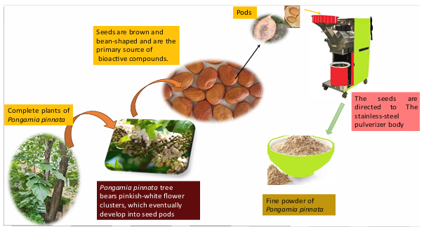
Figure 1: This diagram visually represents the processing of Pongamia pinnata seeds into powdered form through a mechanical process.
FIGURE 2
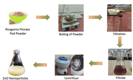
Figure 2: Synthesis schematics of the ZnO nanoparticles
FIGURE 3
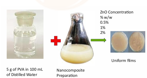
Figure 3: Preparation of PVA-ZnO Nanocomposite Films with Varying Nanoparticle Concentrations
FIGURE 4
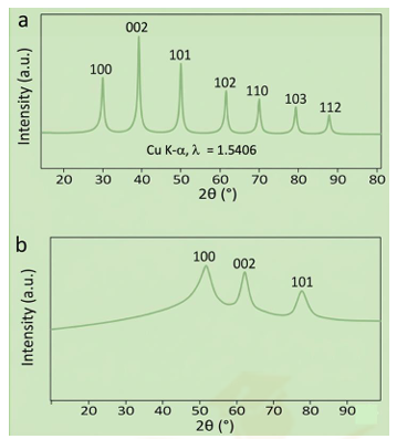
Figure 4: Hexagonal Wurtzite ZnO (a), PVO-ZnO nanocomposite (b)
FIGURE 5
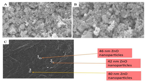
Figure 5: Nanoparticles exhibiting spherical shapes with uniform size at different resolution (a-b), The SEM image shows uniformly distributed, spherical ZnO nanoparticles embedded in the PVA matrix (c).
Tables at a glance
Figures at a glance