Role of Explainable Machine Learning In Early Detection of Osteoporosis
Received Date: October 13, 2025 Accepted Date: October 28, 2025 Published Date: November 16, 2025
doi:10.17303/jmsa.2025.9.104
Citation: Masoumeh Pourzarehsorkhi, Marziyeh Karimi Zarandi, Farzaneh Eskandari (2025) Role of Explanable Machine Learning In Early Detection of Osteoporosis. J Mater sci Appl 9: 1-12
Abstract
Throughout this exploration, analysts utilized artificial neural network (ANN), random forest (RF), and support vector machine (SVM) systems to build a prophetic framework targeted towards customizing treatment tactics for osteoporosis on a personalized level. the framework was crafted to predict alterations in therapy necessary to avert impending fractures due to treatment inefficacy.
Osteoporosis is a progressive skeleton disorder that is often asymptomatic until a fracture occurs, making early detection crucial, yet difficult with traditional diagnostic tools like DXA. ML has great potential in determining early-stage structural bone deterioration, but its clinical adoption is limited. Explainable ML (XML/XAI) sheds light on this issue by showing clinical, demographic, and imaging features that are in line with the model predictions. The goal of this study is to evaluate the use of certain XAI tools (SHAP, LIME, and Grad-CAM) to bolster the effectiveness, practicality, and acceptance in clinical practices of ML algorithms for the early detection of osteoporosis. XAI models build physician trust by allowing them to see the areas of interest that are driving the risk prediction and implementing tailored patient-centered preventive measures.
KEYWORDS: Machine Learning, Osteoporosis, Materials Sciences
Introduction
Osteoporosis, It's a common bone condition that diminishes bone strength, thereby raising the likelihood of fractures. Timely identification and intervention are essential for managing osteoporosis and preventing fractures currently, explainable artificial intelligence (XAI) stands out in the rapid identification of osteoporosis XAI tools can evaluate large sets of bone density images, genetic data, and several other crucial elements they identify trends and initial signs of osteoporosis By providing transparent explanations for its predictions, XAI supports physicians in making knowledgeable choices regarding patients' osteoporosis risk Furthermore, this technology provides opportunities for enhanced comprehension It converts intricate information into comprehensible language Consequently, healthcare providers gain greater confidence in their assessments thus, utilizing XAI, the healthcare experience could change significantly Isn’t that thrilling Innovative methods might transform lives consequently, employing XAI in the detection of osteoporosis can greatly improve patient results in the end, early intervention may result in improved health and reduced fractures Isn’t it incredible how technology can create such an impact With continuous progress, who can predict what’s coming next Furthermore, the partnership between AI and human knowledge can result in significant breakthroughs. Consequently, adopting these advancements is crucial it’s a developing area by joining forces, we can combat osteoporosis more efficiently than ever before the future appears promising, definitely.
However, some specialists argue that relying solely on artificial intelligence algorithms, even those that are understandable, to swiftly detect osteoporosis may overlook the importance of clinical judgment and human expertise. they suggest that although Explainable Artificial Intelligence (XAI) can provide valuable insights, it should be used as an enhancement rather than a replacement for the expertise of medical professionals in diagnosing and managing osteoporosis. Additionally, concerns exist regarding the possible biases in the training datasets of artificial intelligence models, which may lead to incorrect or inappropriate assessments of osteoporosis risk in specific population groups.
Osteoarthritis (OA), a prevalent form of rheumatic disease, is a joint condition that arises with aging. Pain and restricted movement are the primary indicators of OA, with the knee joint typically being the most affected region. In 2019, more than 520 million individuals globally were impacted by OA, as the increasing aging population led to a notable surge in new instances. This is putting increasing pressure on healthcare systems and communities worldwide. In the United States, annual costs from medical expenses and lost income resulting from OA are estimated at 303 billion dollars. At present, no drugs can change the advancement of OA. Recognizing patients with OA at an early stage, or those more likely to deteriorate, may assist in directing treatment choices and result in more focused therapeutic strategies. Currently, X-rays represent the 'gold standard' for diagnosing radiographic osteoarthritis (ROA). Nonetheless, these X-rays are deficient in sensitivity and specifcity concerning alterations in bone and cartilage, resulting in a postponement of prompt clinical intervention [1]. MRI can detect irregularities in bones and soft tissues before radiographic signs appear. Different methods exist for assessing OA abnormalities through MRI; nonetheless, a universally accepted diagnostic standard has yet to be established. Furthermore, Swift recognition is crucial for preventing bone breaks and minimizing adverse impacts on health Innovative tech, such as artificial intelligence, shows promise in rapidly spotting osteoporosis; however, the uncertainty around its frameworks poses a challenge Indeed, timely detection matters greatly; it can save lives and improve overall wellness Moreover, the complexity of these technologies sometimes confuses users, making adoption hard So, while machine learning can revolutionize how we find bone issues, we must address the doubts surrounding it In conclusion, effiective identification strategies are essential; they can lead to healthier futures for many
Artificial intelligence (AI) has produced impressive outcomes in medicine, especially in orthopedics recent research utilizing NHANES data has shown AI's ability to predict OP risk. With ongoing technological progress, it becomes more apparent that AI can exceed the abilities of clinicians in certain situations and may aid in creating personalized therapies. Even with these progressions, a signifi- cant gap remains: the lack of a trustworthy, affordable tool that can be incorporated into hospital environments to identify reduced BMD without needing extra diagnostic tests
Through explainable machine learning (XAI), transparency can be achieved regarding the workings of machine learning algorithms, thus enhancing credibility and practicality in clinical settings. The primary focus of the research is to explore the impact of XAI in the early detection of osteoporosis, highlighting its advantages, obstacles, and future possibilities.
XAI possesses the power to aid healthcare experts in understanding the many elements that impact the chances of osteoporosis in individuals. This might bring about more personalized and effective preventative measures Furthermore, XAI could boost patient trust in machine learning predictions, which ultimately leads to better compliance with recommended osteoporosis prevention strategies. Additionally, XAI plays a crucial role in identifying possible biases in machine learning systems, thereby improving fairness and accuracy in evaluating osteoporosis risks Moreover, XAI can assist in identifying specific traits or biomarkers that strongly indicate osteoporosis this discovery could pave the way for developing sharper and more precise tools for screening, increasing the overall effectiveness of early detection strategies for osteoporosis In this way, XAI transforms the landscape of healthcare by enabling tailored approaches, promoting adherence, and ensuring fair evaluations What a game-changer for osteoporosis management thus, we can see how vital XAI is in advancing our strategies in this critical health area.
Furthermore, transparent machine learning can boost the clarity of prognostic models, allowing healthcare providers to make more knowledgeable judgments concerning osteoporosis risk evaluation [4]. Ultimately, this could pave the way for swifter diagnosis and intervention, ultimately boosting patient results (Hiroshi Akura, Takafumi Nakano, Toshinobu Hayashi, Masashi Takahashi, T. Egawa, 2021). XAI can also aid in crafting more precise fracture risk forecast models by pinpointing the critical risk elements and their relationships.
XAI can enhance the prognosis and intervention of osteoporosis by providing insights into disease progression.
Approximately one in three women and one in five men suffer from Osteoporosis with negatrive consequences like heightened fracture risk and deteriorated health. Conventional diagnostic methods are ommonly used, but they often fail to see microarchaitectural changes. Machine Learning Systems, because of their ability to capture complex nonlinear relationships in imaging and clinical data, have the potential to obviate these challenges. Unfortunately, the opaque nature of these systems as \"black boxes\" has resulted in their stunted clinical usae. Explainable Machine Learning helps overcome this limitation by offering clinical insights into the decision making process of these systems.
Assessment of Osteoporosis
Artificial Intelligence in the realm of medical imaging has made significant strides, thus allowing for earlier detection & intervention. this improvement leads to better outcomes for patients biomedical imaging techniques, driven by AI, present precise diagnostic tools to tackle various medical issues. Moreover, AI-powered imaging systems rapidly analyze medical images, detect patterns, and deliver accurate diagnoses in no time additionally, automation through AI in activities such as image reconstruction signifi- cantly saves time for healthcare workers. As a result, it greatly enhances planning for treatments The effciency gained from these technologies ultimately transforms patient care, allowing doctors to focus more on their patients’ needs In summary, the integration of AI in medical imaging not only streamlines processes but also boosts the quality of care. With every advancement, we witness a shift towards a more effective healthcare system therefore, the future shines brightly with the promise of AI, benefiting both patients & healthcare providers alike.
AI has improved the effciency of biomedical imaging techniques and contributed to the advancement of image-guided interventions and surgeries. throughout surgical procedures, resulting in enhanced precision, exactitude, and patient welfare. To sum up, the assimilation of artificial intelligence in biomedical imaging has triggered a signifi- cant transformation in medical diagnostics and therapy mapping, presenting more precise, effective, and tailored resolutions for healthcare experts and patients. As artificial intelligence advances, we can anticipate additional progressions in medical imaging methodologies, ultimately culminating in enhanced patient care and consequences [7]. Artificial intelligence has further boosted the precision of image interpretation and permitted swifter and more accurate diagnoses. this carries substantial implications for patient care and therapy mapping. Moreover, AI-facilitated imaging has elevated the identification of subtle irregularities in medical images, resulting in earlier diagnosis and therapy. this could potentially lead to improved patient outcomes and survival rates.
Background
Osteoporosis incidents detection occurs primarily through machine learning techniques incorporating either radiological data, data from different databases, or using holistic methodologies. Recent years have witnessed the popularization of various machine learning interpretability techniques primarily aimed at healthcare practitioners, including SHAP, LIME, and Grad-CAM.
Models Training Details
Cutting-edge medical imaging practices play a vital role in the realm of artificial intelligence tools for engineering. Various methodologies such as X-ray imaging, ultrasound, CT scans, MRIs, and PET scans are employed in the domain of medical imaging. Every technique possesses its own set of advantages and hurdles when integrated with AI systems for engineering objectives.
X-ray imaging is widely utilized for scrutinizing mechanical components in machinery and automobiles, and when amalgamated with AI systems, it can heighten precision in pinpointing potential issues.
Ultrasound is imperative for scrutinizing soft tissues within the body and proves to be invaluable in medical diagnostics. AI systems can be educated to scrutinize ultrasound images, aiding healthcare professionals in offering more precise and punctual diagnoses.
CT and MRI produce detailed images of body structures for analyzing irregularities. AI automates interpretation for faster, more accurate problem identification.
PET imaging monitors metabolic processes and detects molecular abnormalities. AI analysis of PET images can identify subtle changes indicating disease progression or treatment efficacy. the combination of imaging methods with AI enhances diagnostics, monitoring, and maintenance in medical and industrial settings. AI integration with X-ray imaging allows efficient, accurate analysis for early issue detection. this integration can lead to quicker, precise diagnoses in medical and industrial environments, improving equipment treatment and maintenance efficacy [10].
Automated systems with artificial intelligence are capable of examining nuclear medicine imaging methods such as SPECT in order to detect subtle changes in tissue functionality or blood flow, resulting in more precise diagnoses and monitoring of medical conditions.
The utilization of AI can help in interpreting sonographic images, enhancing the effectiveness and precision of medical evaluations and prenatal assessments. By incorporating AI into ultrasound imaging, there is the potential for automated image analysis, which leads to quicker and more accurate identification of potential issues or abnormalities.
- AI possesses the power to enhance the evaluation of MRI images. It offers intricate insights into soft tissues, organs, & abnormalities. Thus, these insights can be utilized to formulate more personalized and effective treatment strategies. Moreover, when AI combines with PET imaging techniques, it gives a holistic perspective on metabolic activities this fusion helps in the early spotting and tracking of ailments, notably cancer and brain disorders.
- Furthermore, the blend of AI and PET imaging showcases incredible potential to boost the precision of disease identification. This leads to swift actions and better patient results, especially in battling cancer. Hence, this mixture of AI and PET imaging amplifies the understanding of how diseases develop and how they respond to therapies. Consequently, this ultimately enhances patient care and treatment results In addition, such innovative approaches pave the way for significant advancements in medical practices & methodologies. Not only do they promise better diagnosis, but they also ensure that patients get the attention they truly need. Thus, AI revolutionizes how we perceive and tackle health challenges today. Therefore, continuous progress in this field is crucial. Exciting times ahead, indeed.
Technological Innovations
Mathematical models and algorithms are crucial for scientific inquiry, aiding in exploring complex phenomena, making predictions, and identifying patterns in large datasets. They have significantly impacted our understanding of the natural world and have driven technological advancements with various potential applications.
The collaboration between mathematical models and algorithms has led to advancements in neural networks, with early multilayer perceptron networks having limitations like a superficial structure and limited capacity to represent intricate patterns.
The development of more suitable activation functions like ReLU addressed these limitations and became essential in modern deep learning architectures.
Early multilayer perceptron networks struggled to capture complex patterns, leading to the need for more advanced architectures and training techniques. Enhancements in the functionality of artificial neurons, advanced architectures, and improved training algorithms supported by graphical computational units (GPU) have opened up promising possibilities. The LeNet-5 architecture, created for recognizing handwritten digits, marks a significant milestone for convolutional neural networks (CNNs). These networks, inspired by the visual system of animals, assume that the input represents image data. Current architectures follow a structured sequence of layers, each with specific functions for processing and extracting features from the input data.
The journey begins with the input layer, which receives raw image data, usually in the form of a grid of pixel values, often with three color channels (red, green, blue) for color images. Following the input layer, the network utilizes convolutional layers to extract features. These layers employ various types of convolutional operations to identify local patterns and features in the input data. Initial convolutional layers focus on detecting fundamental features such as edges, corners, and textures. This part examines different methods and algorithms for data engineering and constructing ML pipelines. Our study was performed using Python 3.8.10 on a system equipped with an NVIDIA GeForce GTX 1050 Ti GPU. Libraries in Python such as Pandas, NumPy, SciPy, and Scikit-learn were utilized for data processing, visualizations, and developing machine learning classifiers.
After each convolution operation, activation layers with ReLU activation functions are applied to introduce nonlinearity. These ReLU units help the network learn more intricate patterns and improve its ability to effectively model the data. Subsequently, pooling (subsampling) layers reduce the spatial dimensions of the feature maps while retaining crucial information. Common operations like max pooling and average pooling aid in making the network more resilient to scale and position variations. [14]. Pooling (subsampling) strata might be utilized once more, additional declining the spatial dimensions of the characteristic maps and keeping crucial info. At the tail end of this sequence, subsequent to the network has extracted the most pertinent info from the input data, a distinctive set of vectors are attained, marked by profound features. These, situated deep in the network, distill data into concise, significant forms that are exceedingly discriminative. Or, in different words, after the gradual extraction of info, layer after layer, raw input data is refined into more condensed and theoretical representations that are imbued with semantic meaning, encapsulating crucial features of the input. Profound features can additionally aid alleviate over fitting, a common difficulty in machine learning, as by mastering pertinent representations, they hinder models from memorizing the training data and urge more robust generalization[15]. Another excellent benefit of profound feature extraction pipelines is the potential of utilizing transfer learning techniques. In this instance, a profound feature extraction network formerly effectively developed on one task or dataset might be transferred and fine-tuned to another correlated task, significantly lessening the necessity for large, labeled datasets and hastening up model training. This flexibility is a game changer in myriad applications. Following this extraction front end, proceeding with the processing pipeline and advancing towards the conclusion of the network, completely connected strata are introduced. These strata come after the convolutional and pooling strata and play a pivotal role in feature aggregation and classification. The profound features extracted by the preceding strata are flattened and processed through one or more fully connected strata. In the realm of medical imaging research over the past decade, the exponential growth of imaging data has posed challenges for physicians in efficiently processing images while maintaining accuracy. Fortunately, the exponential growth in computational power has provided a promising foundation for the advancement of AI in medical imaging research [16].
Artificial intelligence has successfully been implemented in radiology to detect abnormalities visible to the naked eye, advancing the field towards a more objective realm of science. Within the realm of Radiation Oncology, AI has been utilized for the precise delineation of tumors and organs, as well as the real-time monitoring of tumor response to treatment.
The notion of "Radiomics" was introduced in the year 2012, entailing the extraction of a multitude of image characteristics from radiographic images. Radiomics is perceived as an innovative strategy for personalized healthcare. Artificial intelligence has displayed significant promise in the realm of medical imaging, instigating conversations about the potential for AI to entirely supplant clinicians.
Immediate challenges facing AI in the medical sphere include the scarcity of high-quality data and con-cerns regarding patient privacy. In the long run, artificial intelligence is anticipated to fall short of human accuracy in medical decision-making due to the subjective nature of healthcare. Ultimately, medical determinations will continue to hinge on human assessment in order to achieve the best possible outcomes for patients [17].
The automatic identification/detection of fiducial markers is necessary for real-time monitoring of the treatment area during administration. Many typical techniques necessitate prior knowledge of the marker characteristics to establish a model. A recently proposed deep learning convolutional neural network (CNN) architecture does not require prior knowledge of marker characteristics or additional training periods to differentiate between cylindrical and irregularly shaped fiducial markers. The system achieved exceptional classification accuracy. Radiomics, an advanced artificial intelligence application in medical imaging research, represents a groundbreaking approach to precision medicine. Radiomics encompasses two key stages. The initial phase involves extracting features. Images from various modalities may be incorporated. Following image segmentation procedures, features are extracted. Common features consist of texture, geometric data, tumor volume, shape, density, pixel intensity, and more. The subsequent stage entails incorporating the extracted features into mathematical models to interpret the tumor's phenotype for forecasting treatment outcomes. Accurate outcome predictions can offer valuable insights for tailoring precise treatment plans. For example, disparate lung cancer patients may exhibit similar histological characteristics and age. However, their tumor images may vary significantly, leading to differing survival durations. By utilizing radiomics to interpret image data, determine the phenotype, and thereby forecast survival times or prognoses before treatment, healthcare providers can select appropriate treatment strategies. This concept is known as personalized or precision medicine. Traditionally, precision medicine relied on biomarkers to predict individual patient outcomes or subtypes, typically through invasive biopsies. In contrast, radiomics does not necessitate invasive procedures. Studies have demonstrated that features extracted solely from CT scans of lung cancer patients are strongly correlated with gene mutations and possess prognostic capabilities. The success of radiomics has the potential to circumvent the risks associated with biopsies and achieve comparable or superior predictive results. [18].
On the policy level, there are increasing concerns on patient privacy. Patient-related health information was protected by tight privacy policies, which limited cross-institution image sharing. Recently, there were several headline news level healthcare data breaches and security attacks. As a result, hospitals are now more than ever concerning about securities and liabilities and have tightened up security and data sharing policies. However, the success implementations of AI needs large amount of data from multiple institutions. How to share images without compromising security is a challenge [19].
The Future of Ai in Medical Imaging Research
Before artificial intelligence (AI) is more widely utilized in medical imaging research, two main challenges need addressing. First, organizing and pre-processing data from various institutions is a key issue. Miotto et al. emphasized the difficulties in summarizing and representing patient data, hindering the widespread adoption of predictive modeling using electronic health records. They introduced an innovative unsupervised deep feature learning approach to create a general patient representation from electronic health record data, facilitating clinical predictive modeling. Although successful in deriving patient representations from a large-scale dataset not tailored to any specific task, handling datasets from multiple institutions poses a more intricate challenge. Variations in procedures and patient cohorts across institutions require meticulous data pre-processing for AI algorithms. In the long run, the question of whether AI can achieve true "intelligence" similar to human capabilities is crucial in determining its potential to replace humans in medical imaging. Unlike purely quantitative tasks, decision-making processes in medical imaging involve knowledge derived from life experiences and philosophical considerations. Achieving human-level behavior in machines presents challenges not only in data collection and algorithm development but also in navigating ethical regulations. The goals for treating osteoporosis are to slow or stop bone loss and to prevent fractures. Your health care provider may recommend:
- Proper nutrition.
- Lifestyle changes.
- Exercise.
- Fall prevention to help prevent fractures.
- Medications.
People facing osteoporosis because of various health issues should work hand in hand with their medical professionals. It’s crucial to identify and tackle the underlying reasons. Changing medication dosages or switching to other therapies may help prevent bone loss for those suffering from osteoporosis. Moreover, patients dealing with conditions that require long-term glucocorticoid treatment can explore drugs approved for preventing or managing osteoporosis. So, it's really important to have open conversations with doctors. They can guide you through many options. For instance, adjusting your current medications could be a game changer. Additionally, considering new medication might provide better outcomes. In fact, understanding the root causes is vital. This way, you can devise an effective plan together. Remember, staying proactive in your treatment is key. It’s not just about managing osteoporosis; it’s about improving your overall health too. Get informed and take charge, because your bones deserve it. Furthermore, look into all available resources. Researching and asking questions leads to better decisions. Always, prioritize your health and communicate any concerns with your healthcare team. They are there to help you navigate this journey. Together, you can find the best solutions for preventing bone deterioration
Research has indicated the efficacy of employing X-ray and CT scans in the identification of fractures, with certain studies achieving notably high AUROCs. Utilizing CNN models has proven to be valuable in the detection of fractures across a range of bodily regions, including the wrist, shoulder, vertebrae, and femoral neck.
Characteristics captured in X-ray imagery, in conjunction with patient information and hospital procedural variables, can be leveraged to distinguish fractures. On occasion, models ascertain fractures indirectly by analyzing associated clinical variables rather than directly relying on image characteristics.
The performance of fracture detection may be impacted by model imbalance, with a greater PRC observed in case-control cohorts exhibiting a higher prevalence of the condition. (The prevalence of 3%).
In particular, in imbalanced tasks such as detecting fractures, data augmentation was attempted in some studies to control the overfitting problem. Some studies have used sampling methods to handle class imbalance. In a recent study, images with data augmentation techniques of generative adversarial networks and digitally reconstructed radiographs from CT showed better performances than those without augmentation (AUROC of 0.92 vs. 0.80, accuracy 86.0%, sensitivity 0.79, specificity 0.90, PPV 0.80, NPV 0.90) [21]. Another recent study reported that the accuracy of fracture detection increased with larger training dataset sizes and mildly improved with augmentation. Consequently, larger studies with optimal augmentation techniques are needed for real-world application of automatic ML-driven detection systems, which may reduce the time and burden of radiologists.
Explainable Machine Learning in Osteoporosis Detection
Feature-Level ExplainabilitySHAP values allow quantification of the impact of each feature including bone mineral density (BMD), age, BMI, vitamin D level, or family history on individualized predictions. This enables clinicians to identify patient-specific risk factors.
Image-Level ExplainabilityGrad-CAM techniques highlight spatial regions in X-ray and CT images that contribute to classification outcomes. Such maps help radiologists validate whether the model focuses on anatomically relevant bone structures.
Model Transparency and Clinical TrustXAI fosters trust by making predictions reproducible and interpretable. Clinicians can understand why a model categorizes a patient as high-risk, enabling easier clinical integration.
Practical Applications
- Early screening in primary care settings
- Automated analysis of DXA and X-ray images
- Risk stratification for populations at high risk
- Personalized prevention and treatment planning
Explainable ML supports:
When used in conjunction with clinical judgment, XAI-enhanced systems reduce false negatives and improve diagnostic reliability
Challenges and Limitations
Despite its advantages, XAI faces challenges including:
- Variability of imaging protocols
- Need for standardized datasets
- Overinterpretation of explanation maps
- Integration with clinical workflow systems
Further research is needed to refine explanation fidelity and develop unified guidelines for evaluating XAI in medical diagnostics.
Conclusion
Uncovering the opaque ML models assists in building trust, transparency, and clinically useful early identification of osteoporosis. XAI broadens physicians' trust in ML and enhances accuracy in diagnosis and tailored patient care. The continued work is needed in real HTML clinical validation. Energizing multilayer XAI solutions is also crucial.
- Kanis JA, et al. (2013) European guidance for the diagnosis and management of osteoporosis in postmenopausal women. Osteoporos. Int. 24: 23–57.
- Wright NC, et al. (2014) The recent prevalence of osteoporosis and low bone mass in the United States based on bone mineral density at the femoral neck or lumbar spine. J. Bone Miner. Res. 29: 2520–26.
- Svedbom A, et al. (2013) Osteoporosis in the European Union: A compendium of country-specific reports. Arch. Osteoporos. 8, 1–218.
- C.D. Lopez, et al. (2021) Using machine learning methods to predict nonhome discharge after elective total shoulder arthroplasty JSES Int.
- P Borbas, et al. (2022) Risk factors for dislocation after reverse total shoulder arthroplasty: a systematic review and meta-analysis Orthopedics.
- Ying H, Guo BW, Wu HJ, Zhu RP, Liu WC, Zhong HF (2023) Using multiple indicators to predict the risk of surgical site infection after ORIF of tibia fractures: a machine learning based study. Front. Cell. Infect. Microbiol.
- Liu WC, et al. (2022) Using machine learning methods to predict bone metastases in breast infiltrating ductal carcinoma patients. Front. Public Health 10: 922510.
- Yu EW, Tsourdi E, Clarke BL, Bauer DC & Drake MT (2020) Osteoporosis management in the era of COVID-19. J. Bone Mineral Res. 35: 1009–13.
- P Pan, et al. (2021) Tumor segmentation in automated whole breast ultrasound using bidirectional LSTM neural network and attention mechanism Ultrasonics.
- KA Walsh, et al. (2021) Performance potential of classical machine learning and deep learning classifiers for isometric upper-body myoelectric control of direction in virtual reality with reduced muscle inputs Biomed. Signal Process. Control.
- N Tsiknakis, et al. (2021) Deep learning for diabetic retinopathy detection and classification based on fundus images: a review Comput. Biol. Med.
- Hong N, et al. (2022) Deep learning-based algorithms to detect vertebral fractures and osteoporosis using lateral spine X-ray radiograph Bone Rep.
- R Wang, J Zhang, B Shan, et al. (2022) XGBoost machine learning algorithm for prediction of outcome in aneurysmal subarachnoid hemorrhage Neuropsychiatr Dis Treat. 18: 659-67.
- S Guan, X Chen, Y Chen, et al. (2022) FOXM1 variant contributes to gefitinib resistance via activating Wnt/β-catenin signal pathway in patients with non-small cell lung cancer Clin Cancer Res. 28: 3770-84.
- S Kim, BR Kim, HD Chae, et al. (2022) Deep Radiomics-based Approach to the Diagnosis of Osteoporosis Using Hip Radiographs. Radiol Artif Intell. 4: e210212.
- YW Jiang, XJ Xu, R Wang, et al. (2022) Radiomics analysis based on lumbar spine CT to detect osteoporosis Eur Radiol: 32: 8019-26.
- BM Touban, MJ Sayegh, J Galina, et al. (2022) Computed tomography measured psoas cross sectional area is associated with bone mineral density measured by dual energy X-ray absorptiometry J Clin Densitom. 25: 592-98.
- BR Bukowski, KP Sandhu, JT Bernatz, et al. (2023) CT required to perform robotic-assisted total hip arthroplasty can identify previously undiagnosed osteoporosis and guide femoral fixation strategy Bone Joint J. 105-B: 254-260
- PL Xiao, CJ Hsu, YG Ma, et al. (2022) Prevalence and treatment rate of osteoporosis in patients undergoing total knee and hip arthroplasty: a systematic review and meta-analysis Arch Osteoporos, 17: 16.
- Z Wang, JE Levin, TB Amen, et al. (2022) Total joint arthroplasty and osteoporosis: looking beyond the joint to bone health J Arthroplasty. 37: 1719-25.
- S Gao, Y Zhao (2023) Quality of life in postmenopausal women with osteoporosis: a systematic review and meta-analysis Qual Life Res. 32: 1551-65
FIGUR 1
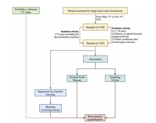
Figure 1 Biomedical Imaging Techniques in AI Systems Engineering Tools
FIGURE 2
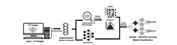
Figure 2: Machine learning model classification
FIGURE 3
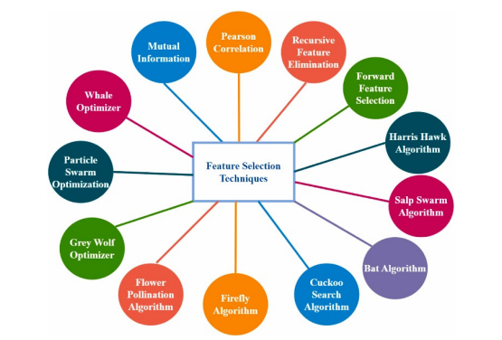
Figure 3: Feature selection techniques
FIGURE 4
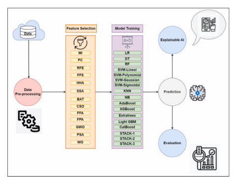
Figures 4
Figures at a glance