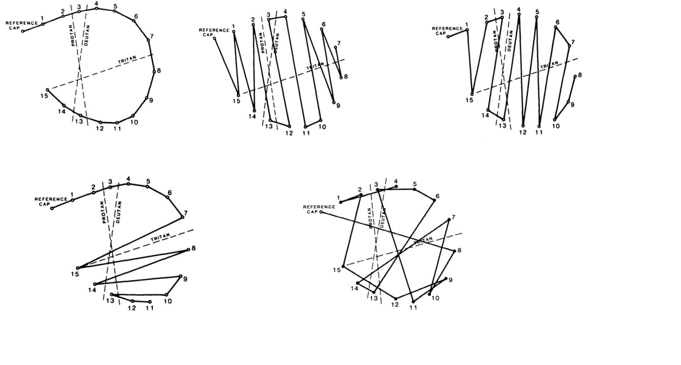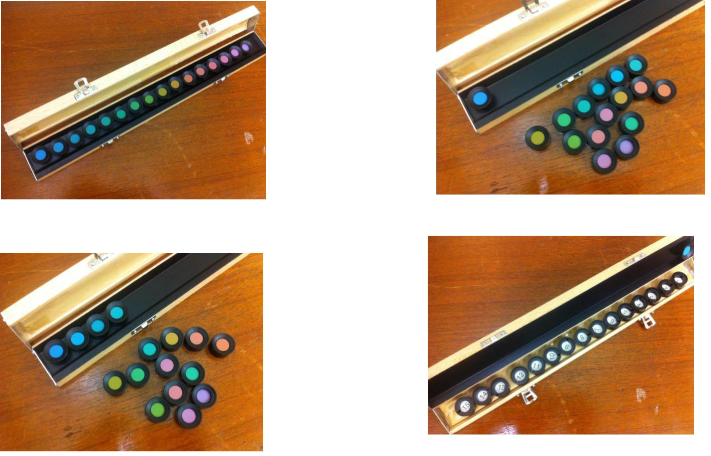
Figure 1.1 Farnsworth D-15 hue test set with 15 coloured caps with a natural hue progression
Figure 1.2 Patients are asked to select a coloured cap that most closely resembled the hue of the reference cap.
Figure 1.3 All 15 caps are arranged in sequence based on the patient's perception of colour
Figure 1.4 After completing the task, the wooden box is flipped over and the corresponding numbers are shown.