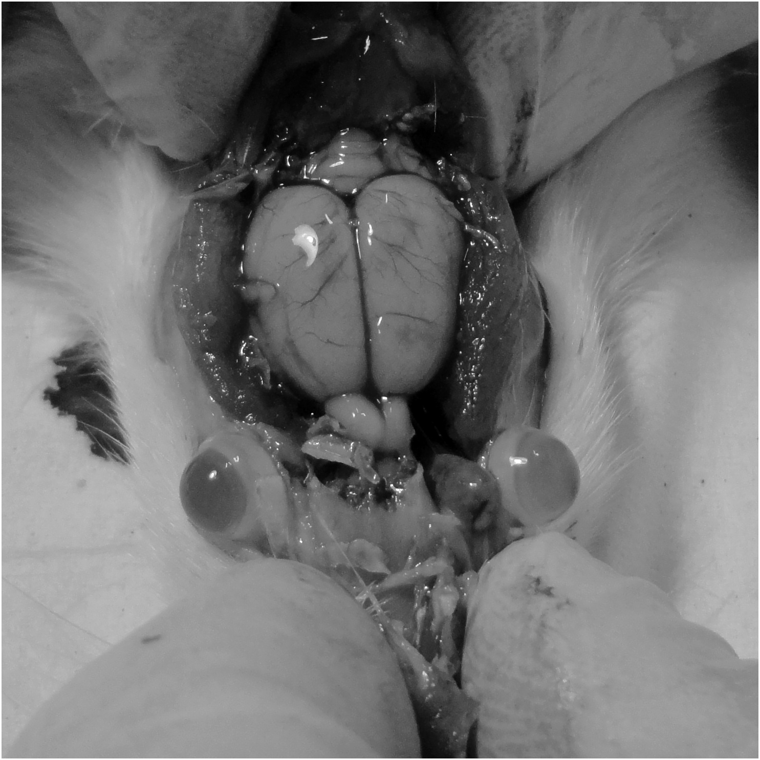
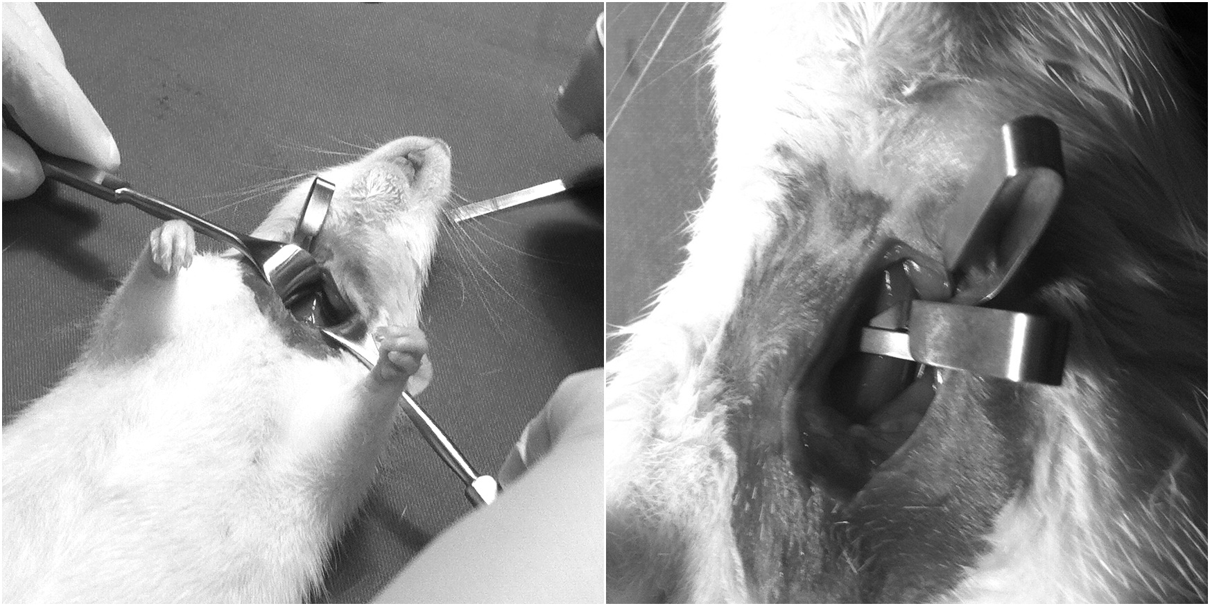
Figure 1 Clamping of bilateral common carotid arteries. Left and then right common carotid arteries were occluded with microsurgical vessel clips within a minute of each other and left in place for 15 minutes to induce cerebral ischemia. The incision was covered with dampened gauze to prevent tissue drying.
Figure 2 Skull cap removal. The skull cap was cut from each orbit, laterally to the rear and reflected forward. Whole brains were removed, cerebellum and olfactory bulbs dissected off for analysis of cerebral tissue.
Figure 3 Coronal slices of cerebrum made using a rat brain matrix. To ensure evenly sliced sections, several blades were positioned in series in every second slot and a single slicing action was employed. Blades were removed individually and neural sections transferred directly to the staining container.
Figure 4 Percentage neural tissue injury per group for males and cyclic and acyclic females. Absorbance values per experimental animal was used to calculate neural tissue injury against absorbance values of animals allocated as controls to each group. Standard error was calculated to be ± 0.619%. Stars indicate significance, males with significantly higher neural tissue injury than both female groups, and acyclic females with significantly higher neural tissue injury than the cyclic female group.