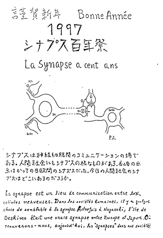
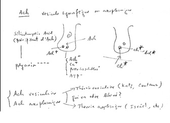
Figure 1 Draft written in French by Tsuji on the two main theories of acetylcholine (Ach) release in the cholinergic synapse. Acetylcholine is released in the synaptic cleft from exocytosis of synaptic vesicles (vesicular theory, favoured by B. Katz and R. Couteaux) or through axoplasmic diffusion (non-vesicular or axoplasmic theory, favoured by M. Israël). Still today, the mechanism of acetylcholine release remains to be clearly elucidated.

Figure 2 Postcard made by Tsuji on the occasion of the 1997 New Year. The year 1997 marked the centenary of the introduction of the concept of the synapse by CS Sherrington (see 10). In the following text, originally written in Japanese and in French, Tsuji compared the synapses to inter-relation places in human societies:"The synapse is a place of communication between nerve cells. In human societies, something is similar to synapse. In the past in Nagasaki, Deshima island was a true synapse between Europa and Japan. Where could we find, today, synapses in our societies?" (from "Le Cerveau au Microscope". (2017). Barbara J-G and Clarac Feds., Hermann, Paris.

Figure 3 AChE activity in a dopaminergic neuron of the rat substantia nigra detected by Tago's method without metallic impregnation, and observed at electron microscopic level: The dark precipitates of DAB oxidized by cupric ferrocyanide are finely localized in the endoplasmic reticulum (ER) and in the nuclear envelop. M: mitochondria, N: nucleus. (x 24,000). (From Tsuji, 1998)

Figure 4 Cytoplasm of a dopaminergic neuron at electron microscopic level. AChE activity detected by Tago's method without metallic impregnation is visible as dark precipitates in the cisternae of the Golgi apparatus (G). M: mitochondria. (x 84,000). (From Tsuji, 1998)

Figure 5 Ultrastructural localization of acetylcholine-like cations in a frog neuromuscular junction at 25 °C (resting state). Point-like precipitates of acetylcholine-like cations are observed in the synaptic vesicles of the nerve terminal (NT). JF: a junctional fold of the synaptic cleft, M: muscle cell.(x 100,000)

Figure 6Ultrastructural localization of acetylcholine-like cations in frog neuromuscular junction at 0°C. The diffusion of acetylcholine-like cations in the synaptic cleft is reduced at low temperature, which enables the visualization of pairs of diffuse precipitates beneath the sites of the active zone of the nerve terminal (NT). M: muscle cell. (x 100,000)

Figure 7 Ionic fixation of acetylcholine-like cations after tyrosine hydroxylase (TH) immunostaining in the substantia nigra of post-mortem human brain. Two nerve endings (N1 and N2) are observed in synaptic contact with a TH-immunoreactive dendritic process of a dopaminergic neuron (TH) stained by dark DAB precipitates. Point-like precipitates of acetylcholine-like cations are present in the synaptic vesicles of N1 and absent in the synaptic vesicles of N2.(x 60,000)

Figure 8Ionic fixation applied to the frog neuromuscular junction after loading with [3 H] choline and brief tetanic stimulation. The cytoplasm of a fibrocyte situated in the vicinity of the neuromuscular junction is intensely labeled with silver grains whereas the nucleus is almost devoid of the autoradiographic label. (x 20,000), (reproduced from the figures of ref. 44).