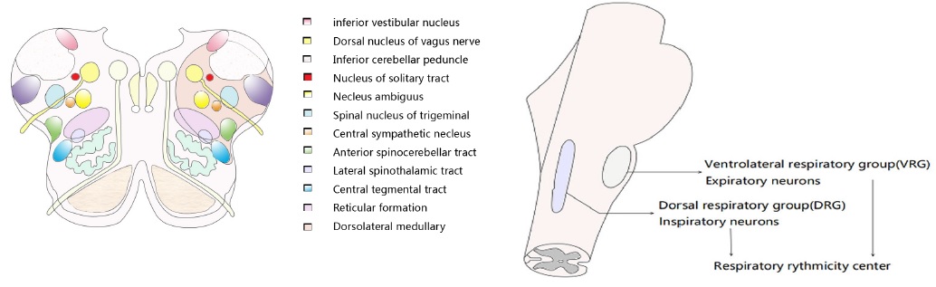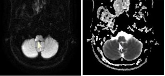
Figure 1 A and B(Patient's brain MRI) : A: Image of craniocerebral DWI cross-sectional scan revealed a signal at the right side of the medulla oblongata(white arrows); B: the corresponding ADC showing a low signal involving pyramidal tracts and reticular structures (white arrows).