A Narrative Study in the Use of Nanoparticles for the Diagnosis and Treatment of Leishmania Species
Received Date: November 08, 2025 Accepted Date: November 17, 2025 Published Date: November 21, 2025
doi:10.17303/jnsm.2025.11.102
Citation: Musafer H. Al-Ardi (2025) A Narrative Study in the Use of Nanoparticles for the Diagnosis and Treatment of Leishmania Species. J Nanotech Smart Mater 11: 1-11
Abstract
Leishmaniasis is a global problem that, so early detection and accurate diagnosis of the parasite are key to comprehensive and effective treatment. Due to their high permeability and absorption properties, nanoparticles have the ability to penetrate cell membranes and bind to nucleic acids. Their superior ability to carry and deliver various drug compounds makes them the first and most effective choice for treating and diagnosing the parasite.
This study aims to survey published articles by Iraqi authors on the diagnosis and treatment of leishmaniasis using nanoparticles.
After searching the Iraqi Academic Journals database, along with other international and Arab databases, a total of twenty five articles and ten theses were reviewed. Following this review, twelve research papers and seven theses were excluded, resulting in the selection of thirteen articles and three theses. Abstracts were then extracted from the selected articles, and pertinent information (the type of nanocomposite used, the concentration levels, and the duration of treatment) were recorded regarding the species of parasites.
Recent studies have used eight types of nanoparticles to treat two species of cutaneous leishmaniasis (L. tropica and L. major) and one species of visceral leishmaniasis (L. donovani). Furthermore, only one study addressed the diagnosis of both types of leishmaniasis (cutaneous and visceral) in blood samples. All analyses were conducted in vitro, except for three, which were conducted in vivo.
Metal nanoparticles, including those made from silver, gold, titanium dioxide, zinc dioxide, magnesium dioxide, and iron dioxide, have demonstrated toxicity and lethality against leishmaniasis. The absence of a standard dosage or treatment duration has prevented comparisons of the effects of these compounds and the determination of which is preferable.
Keywords: Nanoparticles; Leishmaniasis; Treatment; Diagnosis
Introduction
Nanotechnology has recently emerged as a promising field of interdisciplinary research due to its widespread use in various scientific fields. Compounds of silver, gold, zinc, nickel, and other nanoparticles have become increasingly popular due to their exceptional properties, such as high surface-to-mass ratios, quantum structures, and the ability to absorb and transport other compounds (drugs, sensors, and proteins) [1]. Nanoparticles play a role in medical applications due to their unique properties. In a remarkable development, the greener manufacturing of metal oxide nanoparticles has gained significant attention in recent years due to their ease of use, environmental friendliness, accessibility, and non-toxicity. Furthermore, nanoparticles have been tested as antimicrobial materials and for enhancing the shelf life of food products [2]. A diverse group of infectious diseases, known as neglected tropical diseases (NTDs), are prevalent in underserved areas of the world (especially in developing countries) [3]. These diseases are prevalent in tropical and subtropical regions due to poor sanitation, insufficient healthcare, and inadequate or inefficient infrastructure. Currently, more than 20 different types of NTDs are prevalent in 149 countries, affecting approximately 1.4 billion people worldwide [4]. Leishmaniasis is one of the most common NTDs, currently affecting approximately 12 million people worldwide, and 350 million people are at risk in 98 developing countries. Leishmaniasis has recently received more public attention because of its high incidence and the significant morbidity it causes [5]. The London Declaration on NTDs called for the elimination of leishmaniasis as a public health problem by 2020 [6]. Leishmaniasis is caused by an obligate parasite of the genus [7]. There are approximately 51 species of these parasites, of which 21 are pathogenic and cause leishmaniasis, including [7, 8]. Leishmania parasites exist in two forms: a round, non-flagellate form and an elongated, flagellate form. The round parasite is small and non-motile, while the elongated form can move with the help of flagella [9]. Transmission of Leishmania occurs when a sand fly sucks blood from an infected person (human or animal) [10]. Metamorphosis of the parasite occurs when it changes from the flagellate stage to the pro-flagellate stage, and it takes about 4-25 days [11]. The disease results in ulcers and also affects other organs of the body [12].
Methods
Articles on the use of nanoparticles of various compounds in the diagnosis and treatment of leishmaniasis in Iraq, which had previously been published in Iraqi academic journals and other international and Arab databases (Scientific Researcher, Pumped, Soci), and Iraqi university websites, were gathered. The following keywords were used in the search: "nanoparticles against leishmaniasis," "nanoparticles," "leishmaniasis treatment," and "use of nanoparticles in the diagnosis of leishmaniasis" in both Arabic and English.
During the search, we identified twenty-five research papers authored by Iraqi researchers and ten scientific dissertations written in either Arabic or English, all of which addressed the research topic. In the first stage, the research papers were selected based on their titles and then further examined by reading the abstracts and keywords. Papers that did not meet the established criteria were excluded.
In the second phase, we reviewed the research methods to ensure they conformed to the research criteria. As a result, some papers were excluded (Figure 1). Specifically, twelve research papers were eliminated for not adhering to the criteria: seven were co-authored with non-Iraqi researchers, three were previous reference studies, and two focused on the Histo- pathological effect of nanocomposites on the host body. Additionally, seven scientific theses were excluded due to insufficient conclusions. Ultimately, thirteen articles and three scientific theses were approved for inclusion in the study.
Results
From thirteen articles and three scientific dissertations by Iraqi authors that addressed the treatment and diagnosis of leishmaniasis in Iraq, the following conclusions were drawn:
Recent articles have used eight types of nanoparticles to treat two species of cutaneous leishmaniasis (L. tropica and L. major) and one species of visceral leishmaniasis (L. donovani). On the other hand, only one study addressed the diagnosis of both types of leishmaniasis (cutaneous and visceral) in blood samples. All experiments were conducted in vitro, except for three that were conducted in vivo (Table 1).
The extracted studies used different concentrations of nanoparticles and varied widely in their time periods, with some lasting minutes and most lasting hours to days. All nanoparticles used were highly inhibitory at the average concentration and for the longest time periods. In only one study did the researcher indicate that treatment with nanoparticles (magnesium oxide) was ineffective (Table 2).
Discussion
The field of using nanoparticles in the treatment of pathogens is new. It is undergoing rapid development to discover the properties of these particles and their superior penetration and ability to reach targets or carry various therapeutic or diagnostic molecules. Therefore, the absence of many articles in this field may be due to its novelty. In this survey, the recent articles have utilised nanoparticles of various materials and concentrations, even for a single substance; this makes comparison or judgment impossible, as they cannot be included in a statistical analysis that yields realistic results.
Titanium dioxide (TiO2) is a non-toxic, water-insoluble material with high stability and excellent catalytic performance. Other properties of TiO2 include a high specific surface area, high crystallinity, and a high concentration of surface hydroxyl [29]. The specific surface area is a parameter in photocatalytic efficiency, and a higher specific surface area is usually associated with lower catalyst crystallinity. One of the most important applications of titanium dioxide is its use in photocatalytic processes, particularly when ultraviolet or infrared light is used as a catalyst [30].
The effect of titanium dioxide nanoparticles (TiO2NPs) depends on their particle size. The smaller the particle size, the greater the accumulation on the surface of cells, which causes oxidative stress, greater toxicity to cells and destroys microorganisms on which the nanoparticles act. In addition, they can strongly bind sulfur and phosphorus, compounds found in DNA or that can accumulate significantly within mitochondria, and impair their function [31]. Another mechanism of action of nanomaterials is the release of ions that contribute to cell death by producing large amounts of reactive oxygen species (ROS). ROS generation within metal nanoparticles is an apoptosis mechanism caused by nanoparticle-induced oxidative stress [32]. Since mitochondria are one of the main target organs for oxidative stress, high levels of nanoparticles can cause membrane phospholipid decomposition and induce mitochondrial membrane depolarisation. Various metal oxides mediate ROS nanoparticles, leading to cell death by disrupting mitochondrial function [33].
The remarkable properties of magnesium nanoparticles have made them a good competitor to their silver counterpart. Magnesium oxide nanoparticles are very stable, biocompatible, and very effective antibacterial agents [34]. MgONPs have very effective bactericidal properties; they can lead to the complete elimination of pathogenic microbes [35]. MgONPs damage the cell membrane and cause lipid peroxidation, leading to leakage of intracellular contents [36].
By increasing the concentration of MgONPs, their inhibitory effectiveness can be enhanced. Furthermore, it has been observed that their small size improves their antibacterial properties (both Gram-positive and Gram-negative bacteria). Increasing the surface area exposes more active sites on the nanoparticles' surfaces, thus enhancing their antibacterial properties [37]. MgONPs have demonstrated antibacterial activity against Leishmania species. They showed increased inhibition efficiency against the flagellate stages of L. major [38] and L. tropica [39] after increasing the concentration of the MgONP nanoparticle solution.
Silver oxide nanoparticles have unique chemical and physical properties attributed to their nanoscale structure, making them a new and effective antibiotic with a high antibacterial effect. They work, like other nanoparticles, through the mechanism of oxidative stress. It has been suggested that silver ions play a role as a catalyst in cellular oxidation processes. This catalysis facilitates the formation of a disulphide bond (R-S-S-R) by promoting the reaction between oxygen molecules and hydrogen within the cell. As a result, water is released, which can lead to cell rupture and apoptosis [40]. Silver nanoparticles can also bind silver to sulphur groups (-SH) in enzymes, thus becoming active [41]. Silver nanoparticles can create irregular holes in the outer membrane of the cell and change its permeability. Silver ions also enter the cell and interfere with the bacterial cell, leading to the loss of important molecules for the cell and its death [42]. Silver ion nanoparticles provide an additional bactericidal effect by affecting the acid molecule. Nuclear degradation and loss of the ability to replicate. These molecules can bind to purine and pyrimidine bases in DNA, leading to its disintegration. Ag2O2 NPs have been shown to damage the DNA of Escherichia coli and terminate the cell cycle in the second growth phase (G2/M division) due to DNA damage [43].
Carbon nanotubes consist of graphite sheets rolled into a cylindrical shape. The size and surface properties of the nanoparticles are the main factors affecting their circulation time, bio- distribution, and cellular uptake [44]. In addition, carbon nanotubes are able to cross cell membranes and transport peptides, proteins, and nucleic acids into cells, making them useful as drug delivery vehicles for intracellular targets. Studies have shown that these molecules can be absorbed by lymphocytes and macrophages without affecting cell viability [45].
The properties of carbon nanoparticles make them suitable for various potential applications, including therapeutic applications. The effects of carbon nanoparticles on parasite strains are due to their solubility, size, and shape. The poor solubility of carbon nanoparticles in water leads to aggregation, thereby reducing penetration into cells. Furthermore, the effects of carbon nanoparticles may be reduced due to the reduction of the binding sites between the nanoparticles and the cell surface [46]. On the other hand, these nanoparticles can cross the cell membrane, interact with various compounds within the cell, and prevent cell division. Therefore, the large surface area per unit volume and the high ratio of atoms in the different surface layers make carbon nanoparticles more active in treating diseases [47].
Gold nanoparticles are among the most widely used and studied nanoparticles. Gold is one of the most stable metals, making it the most commonly used in scientific research and study. The production and synthesis of gold nanoparticles follows a top-down process. In this process, gold salts are reduced in the presence of stabilising agents, which prevent the gold particles from clumping together [48]. Gold nanoparticles (AuNPs) are being explored as promising anti-leishmanial agents due to their ability to bind to multiple sites (due to their large surface area) on the cell membrane and their ability to generate ROS. Gold compounds have been reported to inhibit trypanothione reductase, an enzyme in redox metabolism in Leishmania [49].
Gold nanoparticles also serve as binding sites for anti-leishmanial drugs on their surface, thereby enhancing their therapeutic efficacy. Gold nanoparticles exhibit significant activity against Leishmania by silencing the gp63 gene, followed by parasite killing after the short nucleotide chain of the antibody is bound to the drug molecules [50]. It has also been suggested that direct adhesion of nanoparticles to the cell surface disrupts the cell membrane, and the nanocomposite damages proteins and organelles after entering the cell. It also stimulates the release of antioxidants by macrophages in the absence of oxygen, and can accumulate within macrophages, reducing cell proliferation and stimulating inflammation body activation in macrophages, enhancing the inflammatory response [51].
Zinc oxide nanoparticles (ZnO NPs) are one of five zinc compounds registered by the USA Food and Drug Administration as generally safe. These nanoparticles are effective in inhibiting the growth and reproduction of microorganisms, and have anti-bacterial effects on both Gram-negative and Gram-positive bacteria such as Escherichia coli, Pseudomonas aeruginosa, and Staphylococcus aureus [52].
ZnO NPs possess dose-dependent anti-leishmanial activities. Cell viability and proliferation were observed to decrease after treatment with nanoparticles. It was suggested that the nanoparticles destroy mitochondrial enzymes and cell cycle proteins [53]. Furthermore, other studies conducted to investigate the efficacy of zinc sulphate on L. tropica and L. major found that these particles had an inhibitory effect on highly virulent enzymes and carbohydrate enzymes, and the most important regulatory enzyme in the glycolysis pathway was inhibited [54].
An ideal drug delivery system should possess two important elements: controlled and targeted delivery. In this regard, ZnO NPs have emerged as potential and effective drug delivery systems, with which drugs have the optimal concentration at which they are beneficial [55]. Therefore, in designing ZnO NPs, the main goal is to control the particle size and surface properties to achieve controlled release of the pharmacologically active agent at a specific site at the optimal therapeutic rate within the dosage regimen. Due to their extremely small and controllable size, NPs can easily penetrate body cells and, more importantly, they exhibit high interaction with microbial biological systems [56].
Conclusion
Various metal nanoparticles exhibit anti-leishmanial properties that depend on the concentration and duration of their administration, whether in vivo or in vitro, primarily by inducing oxidative stress through the production of reactive oxygen species. Metal nanoparticles, including nanoparticles made of silver, gold, titanium dioxide, zinc dioxide, magnesium dioxide, and iron dioxide, have demonstrated toxicity and thus anti-leishmanial effects. The lack of adherence to a standard dose or treatment duration has prevented comparisons of these compounds and the determination of which is preferable.
Ethics approvals
This article does not contain any studies involving animals performed by any of the authors.
Disclosure and conflict of interest
The authors declare that they have no conflicts of interest.
Acknowledgement
This work was supported by corresponding author “Musafer Al-Ardi"
- P Rai Dhirendra (2021) "A review on concept of nanotechnology in veterinary medicine," ES. Food & Agroforestry. 4: 28-60.
- P Rai Dhirendra (2022) "A review on aspects of nanotechnology in food science and animal nutrition." ES Food & Agroforestry. 8: 12-46.
- S Srivastava, P Shankar, J Mishra, S Singh (2016) "Possibilities and challenges for developing a successful vaccine for leishmaniasis”. Parasit &Vectors. 9: 277.
- World Health Organization (2019) "Global update on implementation of preventive chemotherapy against neglected tropical diseases in 2018–Actualisation à l’échelle mondiale des informations relatives à la chimioprévention des maladies tropicales négligées en 2018,” Wkly. Epidemiol. Rec. 94: 425-38.
- J Alvar, ID Vélez, C Bern, M Herrero, P Desjeux, J Cano, J Jannin, M den Boer (2012) “Leishmaniasis worldwide and global estimates of its incidence," PLoS ONE. 7: e35671.
- World Health Organization (2012) "Uniting to combat neglected tropical diseases in London Declaration on Neglected Tropical Diseases," NTDs: Haywards Health, UK.
- E. Handman, "Cell biology of Leishmania (1999) "Advances in Parasitology. 44: 1–39.
- M Vannier-Santos, A Martiny, W d Souza (2002) "Cell biology of Leishmania spp. invading and evading," Curr. Pharm. Des. 8: 297–318.
- J Alexander, A Satoskar, D Russell (1999) "Leishmania species: Models of intracellular parasitism," J. Cell Sci. 112: 2993–3002.
- PA Bates (2007) "Transmission of Leishmania metacyclic promastigotes by phlebotomine sand flies." Int. J. Parasitol. 37: 1097–106.
- F Bailey, K Mondragon-Shem, P Hotez, JA Ruiz-Postigo, W Al-Salem, "Acosta-Serrano, A Molyneux, (2017). A new perspective on cutaneous leishmaniasis implications for global prevalence and burden of disease estimates," PLoS Negl. Trop. Dis. 11: e0005739.
- K Singh, G Garg, V Ali, (2016) "Current Therapeutics, Their Problems and Thiol Metabolism as Potential Drug Targets in Leishmaniasis." Curr. Drug Metab. 17: 897–919.
- RA Al-Janaby, (2022) "Effect of Titanium Dioxide Nanoparticles (TiO2NPs) on Leishmania parasite in vitro." Wasit Journal for Pure sciences. 1: 78-85.
- H Esmail (2022) "In Vitro Study of Ag2O, MgO Nanoparticles and Sb Drug Cytotoxic Effects on Leishmania donovani", Journal of Biotechnology Research Center. 16: 39-49.
- ST Mohammed (2017) "Effect of Silver Nanoparticles Synthesis from Sphingomonas paucimobilis in Leishmania donovani in vivo and in vitro," International Journal of Chem.Tech. Res. 10: 1028-37.
- RF Al-Saeedi, EJ Saheb, (2017).”Effect of silver nanoparticles on macrophage cytotoxicity upon exposure to Leishmania tropica in vitro”. Iraqi Journal of Science. 2017: 1419-27.
- MT Hannoun (2022). "A therapeutic attempt with silver nanoparticles and the extract of the field horsetail plant Equisetum arvense and the burdock Arctium lappa L for the infection of Leishmania tropica skin disease in the city of Samarra," Master's Thesis, Samarra University, Faculty of Education. 123.
- KH Zghair, E Saheb, B AL-Qadhi (2016) "Antiparasitic Effect of Carbon-nanotubes on Leishmania donovani in vitro," Iraqi Journal of Science. 57: 2641.
- A Taha, A Faisal, A Shaimaa, M Al-Halbosiy (2021) "Antimicrobial Activity of Locally Synthesized Carbon Nanosphere on Some Pathogenic Species of Bacteria and Parasites," Baghdad Science Journal. 18: 1-6.
- N Abdu al Aziz, MA Abudlrahman, AS Mohammed (2022) "Evolution of gold nanoparticles as a drug against cutaneous leishmaniasis in vitro," Samarra Journal of Pure and Applied Science. 4: 48-56.
- A Marhoon (2023) "Evaluation of the Efficacy of Sodium Chloride Nanoparticles on the Vitality of Leishmania Major (in vitro)," Archives of Razi Institute. 78: 627.
- AT Enad, KH Zghair (2016) "Cytotoxic Effect of ZnO Nanoparticles on the Viability of Leishmania donovani Promastigotes in vitro," Iraqi Journal of Science. 57: 2811-17.
- MM Salih, KH Zghair (2017) "The Cytotoxic Effect of Zno Nps against the Intracellular Amastigotes of Leishmania Donovani in Vitro," Iraqi Journal of Science. 58: 2285-90.
- AG Meaad, N Ban (2017) "The Effect of Zinc oxide Nanoparticles (ZnO NPs) on the Viability of Leishmania tropic In Vitro," Iraqi Journal of Science. 58: 600-10.
- JA Mohammed, SJ Mushattat, AJ AL-Arid (2024) "The protective role of nanoextract Origanum majorana leaves and histological changes in the lungs of albino rats treated with Leishmania donovani," Al-Kunooze Scientific Journal. 8: 141-50.
- C Hano, BH Abbasi (2021) "Plant-based green synthesis of nanoparticles: Production, characterization and applications," Biomolecules. 12: 31.
- WS Latif (2021) Therapeutic effect of Neem Alcoholic Extract and Zinc Nanoparticles on Cutaneous Leishmaniasis Parasite. Master's Thesis, University of Tikrit, Faculty of Veterinary Medicine. 112.
- AM Barghash (2022). Biological synthesis of iron oxide nanoparticles using the aqueous extract of cream fruit and testing of the inhibitory effect against the anterior whip of Leishmania tropica. Master's Thesis, University of Kirkuk, Faculty of Science. 124.
- MH Al-Ardi (2022) "Rapid diagnosis of leishmania spp. in blood samples using gold nanoparticles," Iraqi Journal of Veterinary Sciences. 36: 587-90.
- E Dolat, S Salarabadi, P Layegh, M Jaafari, S Sazgarnia, A Sazgarnia (2020) "The effect of UV radiation in the presence of TiO2-NPs on Leishmania major promastigotes," Biochimica et Biophysica Acta (BBA)-General Subjects. 1864: 129-238.
- A Sepúlveda, A Velásquez, I Linares, L de Almeida, C Fontana, C Garcia, M Graminha (2020) "Efficacy of photodynamic therapy using TiO2 nanoparticles doped with Zn and hypericin in the treatment of cutaneous Leishmaniasis caused by Leishmania amazonensis," Photodiagnosis and Photodynamic Therapy. 30: 101- 16.
- RA Abd-Algany (2022) "Effect of Titanium Dioxide Nanoparticles (TiO2NPs) on Leishmania parasite in vitro." Wasit Journal for Pure sciences. 1: 78-85.
- TS Bologna, ID Ferrato de Sousa, JR Neto, TA Nunes, BS Matos (2024) "Antiprotozoal, Anthelmintic and Antivectorial Activities of Titanium Dioxide Nanoparticles," Current Medicinal Chemistry.
- RO Guerra, JR Neto, T Martins, TS de-Assunção, VR Junior (2022) "Metallic nanoparticles: a new frontier in the fight against leishmaniasis," Current Medicinal Chemistry. 29: 4547-73.
- KM Salman, CG Renuka (2023) "Modied sol-gel technique for the synthesis of pure MgO and ZnO nanoparticles to study structural and optical properties for optoelectronic applications," Materials Today.
- IK Abbas, KA Adim (2023) "Synthesis and characterization of magnesium oxide nanoparticles by atmospheric non-thermal plasma jet," Kuwait Journal of Science. 50: 223-30.
- P Yadav, R Saini, A Bhaduri (2023) "Facile synthesis of MgO nanoparticles for effective degradation of organic dyes," Environmental Science and Pollution Research. 30: 1439-53.
- RO Guerra, JR Neto, T Martins, TS Farnesi (2022) "Metallic nanoparticles: a new frontier in the fight against leishmaniasis," Current Medicinal Chemistry. 29: 4547-73.
- S Mohtasebi, M Mohebali, S Elikaee, B Akhoundi, AR Foroushani (2019) " In vitro and in vivo anti-parasitic activity of biogenic antimony sulfide nanoparticles on Leishmania major ," Parasitology research.118: 2669- 78.
- S Sheema, Q Jamal, S Zafar (2024) " Leishmanicidal and Anti-Microbial Effects of MgO–ZnO Bimetallic Nanocomposites, Fabricated with Curcuma zedoaria Oil," Journal of Inorganic and Organometallic Polymers and Materials. 30: 1-2.
- QA Majeed, AF Shater, AD Alanazi (2023) "Green Synthesis, Characterization, and Antileishmanial Activity of the Silver Nanoparticles Alone and Along with Meglumine Antimoniate against Leishmania major Infection,” Iranian Journal of Parasitology. 18: 535.
- A Ebrahimzadeh, M Karamian, E Alemzadeh, R. Solgi, E. Alemzadeh, S. Mortazavi-Derazkola,(2024). "Investigation of therapeutic potential in murine cutaneous leishmaniasis and antibacterial using biosynthesized silver nanoparticles using extract of Crocus sativus petals," Biomass Conversion and Biorefinery. 14: 4485- 96.
- F Sharifi, N Mohamadi, R Tavakoli Oliaee, I Sharifi, M Doostmohammadi, S Soltanian, F Sharififar (2023) "The potential effect of silver nanoparticles synthesized with Coffea arabica green seeds on Leishmania major proliferation, cytotoxicity activity, and cytokines expression level," Journal of Parasitic Diseases. 47: 131-39.
- M Shakeel, MH Kiani, HS Sarwar, S Akhtar, A Rauf, et al. (2023) "Emulgel-loaded mannosylated thiolated chitosan-coated silver nanoparticles for the treatment of cutaneous leishmaniasis," International Journal of Biological Macromolecules. 227: 1293-304.
- H Liu, J Chen, S Qiao, W Zhang (2021) "Carbon-Based Nanomaterials for Bone and Cartilage Regeneration: A Review," ACS Biomater Sci. Eng. 7: 4718−35.
- M Heidari‐Kharaji, S Guerra, RP Puneiad (2024) "Effects of Amphotericin B‐Conjugated Functionalized Carbon Nanoparticles in the Treatment of Cutaneous Leishmaniasis," Parasite Immunology. 46: e13068.
- A Singh, S Sharma, G Yadagiri, S Parvez, R. Gupta, et al. (2020) "Sensible graphene oxide differentiates macrophages and Leishmania: a bio-nano interplay in attenuating intracellular parasite," RSC advances. 10: 27502-511.
- K Dasauni, D Bisht, TK Nailwal (2021) "Novel nanotechnology-based approaches in the treatment of leishmaniasis. In Pathogenesis, Treatment and Prevention of Leishmaniasis," Academic Press. 125-40.
- SM Amini, R Hadighi, M Najm, M Alipour, H Hasanpour, M Vosoogh, A Vosough, M Hajizadeh, A Badirzadeh (2023) "The therapeutic effects of curcumin-coated gold nanoparticle against leishmania major causative agent of zoonotic cutaneous leishmaniasis (ZCL): an in vitro and in vivo study," Current Microbiology. 80: 104.
- MY Want, P Yadav, R Khan, G Chouhan, M Islamuddin, et al. (2021) "Critical antileishmanial in vitro effects of highly examined gold nanoparticles," International journal of nanomedicine. 28: 7285-95.
- K Sharma, J Shah, S Singh, S Sengupta (2024) "Development of Amphotericin B Decorated Gold Nanoparticles as a Promising Antileishmanial Nanoconjugate," ACS Applied Bio Materials. 7: 6239- 48.
- MT Elhefny, NE Mostafa, EA Mohamed, RM Abdelhameed, MH Sarhan, HS Moawad (2021) "Role of nanoparticles in diagnosis and management of parasitic diseases," The Egyptian Journal of Hospital Medicine. 86: 85-99.
- C Khatana, A Kumar, MW Alruways, N Khan, N Thakur, D Kumar, A Kumari (2021) "Antibacterial potential of zinc oxide nanoparticles synthesized using Aloe vera (L.) Burm. F: A Green approach to combat drug resistance, J. Pure Appl. Microbiol. 15: 1907-14.
- F Saleh, F Kheirandish, M Abbasi, F Ahmadpour, S Veiskarami, A Mirderikvand (2024) Comparison of biosynthetic zinc oxide nanoparticle and glucantime cytotoxic effects on Leishmania major ," Journal of Basic Microbiology. 64: 230- 43.
- M Aghaei Aghaei, S Kouhiyan, M Shahmoradi, Z Hejazi (2024) "The Interaction of Zinc as an Essential Trace Element with Leishmania Parasites: A Systematic Review," Advanced Biomedical Research. 1: 73.
- A Karimipour-Saryazdi, MM Jafari, R Omidi, F Ghaffarifar, SH Sadeghi (2022) "Anti-leishmania Effect of Magnesium Oxide Nanoparticles on Leishmania tropica/infantum and Leishmania Infected Macrophages," International Journal of Enteric Pathogens. 10: 144-54.
FIGURE 1
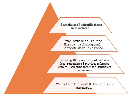
Figure 1: Excluded and Included Research Papers.
Tables at a glance
Figures at a glance