Nanocellulose from Heart-of-Peach Palm Residue: Preparation and Characterization
Received Date: November 15, 2021 Accepted Date: December 15, 2021 Published Date: December 17, 2021
doi: 10.17303/jnsm.2021.7.102
Citation: Cristiane Vieira Helm (2021) Nanocellulose from Heart-of-Peach Palm Residue: Preparation and Characterization. J Nanotech Smart Mater 7: 1-17
Abstract
Increasing use of nano materials and the need to find new sources to produce nanofibrils have led many researchers for preparing and characterizing nano cellulose from agricultural sources. Accordingly, the objectives of this study are preparation and characterization of nanofibrils of cellulose from heart-of-peach palm residue. This paper presents the preparation of nanofibrils from heart-of-peach palm residue by mechanical fibrillation of bleached cellulose pulp. Characterization includes their dimensions, chemical constituents and toxicity. The nanofibrils produced in gel form contained about 97.8g/100g of moisture and 2.2 g/100g of total fiber content. No toxicity was observed to the brine shrimp Artemia salina up to 50 g/L of fibrils indicating they are not toxic to such an animal. It is hoped these results will encourage the search for other nano-structured materials that is non-toxic to the environment.
Keywords: Heart-of-Peach Palm Residue; Bleached pulp; Mechanical fibrillation; Nanostructure; Chemical composition; Toxicity; Brine shrimp. Artemia salina
Introduction
It is well known that extensive research and use of nano materials along with finding new sources for producing nano fibrils have been on the rise. Also, there are many methods to prepare nano cellulose particularly based on bio-resources. It is also reported that there has been continuous developments in the nanotechnology applications in food sector during the last decade or so particularly into the value-added chain of food processing and manufacturing [1-3]. Besides, nano structured materials can be added as thickener or as source of fibers to control intestinal transit or decrease cholesterol, glycaemia and others biochemical parameters. It is in this context complete characterization of nano materials including aspects of toxicology regarding living organisms is needed, when nano fibers are produced from various sources, especially from biomass and when these are used as an additive even in animal feeding with its effective role in various aspects including toxicity and risk of environmental pollution.
Nanotechnology has attracted the attention of scientists in many fields because of the unique characteristics of nanomaterials that are not possible with conventional materials as reported. One of the largest sectors that has benefitted from nanotechnology is the food industry, with the development of functional materials, packaging, additives and others [4-10]. This is due to the unique characteristics of nanomaterials that are not possible with conventional materials. Although a good deal of research has been carried out on nanomaterials by physicists and chemists, resulting in a large number of published reports on the characterization of such materials, legislation on the use of these materials in products such as cosmetics and foods is yet to be formulated [7]. Nevertheless, due to the great potential of nanotechnology for innovations, there have been some reviews on the use of nanotechnology in the food sector such as in packaging, additives and others, including the implications of biological aspects and potential toxicity [5, 9-13]. There have been continuous developments in nanotechnology with even some research groups in various institutions including some private industries have initiated research programs to explore the wide scope of nanotechnology into the value-added chain of food processing and manufacturing [7]. In fact, possibility for the use of silver nano particles as food additives in animal feeding is reported [10] mentioning some caution to be exercised while doing so particularly with respect to environmental pollution besides the toxic effect. This is true even in the case of nanomaterials prepared from biomass and hence characterization of these materials to overcome the risk of environmental pollution is needed particularly when these are to be used for different purposes.Nanofibers can be produced from various sources, particularly from biomass [14] with a number of published reports about their possible use in food sector [7, 9, 11-17]. Production of nano-crystalline cellulose materials from lignocellulosic biomass is reported [15]. This report also highlighted their technological developments and applications and the challenges and future opportunities as well as obstacles for the wide use of these materials. Recently, microcrystalline cellulose (MCC) from totally chlorine-free pulp from oil palm fruit has been successfully isolated using acid hydrolysis [16]. This material showed good thermal stability with potential for a range of applications, including green nano bio-composites and as pharmaceutical tableting material. Biomass is as source has been used to produce cellulose nanofibrils in the form of gel by first homogenizing the material in a high-pressure homogenizer [16-19]. Fibers in this gel have been reported to be of nano-size in diameter but micro-size in length [14].
Abdul Kalil et al [18] have reviewed the production and modfication of nano fibrillated cellulose from biomass using various mechanical processes, stressing the increased attention being given to these materials by pointing out that due to unique characteristics of biomass such as its renewability, low density, high mechanical properties, biodegradability and low cost, increased attention being given to its use in the preparation of nano materials.
Among various biomass materials used as sources of nanofibrils, peach palm trees (Bactris gasipaes) are one of the potential candidates, particularly the heart-of-palm, a widely cultivated plant in Brazil as well as Central America with Brazil representing more than 50% of the whole heart-of-the-peach palm product marketed legally in the world. This culture has been introduced in the South and Southeast Regions of Brazil to avoid exploitation of heart-of-palm from the native juçara palm (Euterpe edulis) [called as ‘palmito’ in Brazil], an endangered species in the Mata Atlântica rainforest.
In the case of heart of peach palm, just one fifth of the inner part of the stem tip can be used as human food [20], while about 70% of the stem is residue [21]. On the positive side, this residue shows great potential as a supplement to add fibrils to animal feed, which promotes several beneficial effects in the animal’s metabolism. This shows a great potential as supplement of fibrils in the animal feeding promoting several beneficial effects in the animal metabolism. Accordingly, this material was chosen to prepare the nanocellulose in this study.
There are some published papers mainly dealing with the characterization of peach palm and processing of nanocellulose from this plant material [22-24]. One of these [22] deals particularly with the chemical composition and the morphology of various parts of peach palm as well as those of sheaths and stem (the processed by-products) flours and other aspects, while the other deals with fractionation of the polysaccharides present in the peach palm by-product flours and other aspects [23]. The last one [24] presents the preparation of nanocellulose directly from the fibers of this material without any normally used previous pulping process during the preparation of nanocellulose from lignocellulosic materials, but using different chemical delignification protocols followed by mechanical disintegration in two different stages. Thus obtained nanocellulose is characterized in respect of chemical composition, thermal properties, crystalline structure and morphologies with the purpose of finding possible future use of this material. It is reported that just one fifth of the inner part of the stem tip of heart of peach can be used as human food [20], while about 70% of the stem is residue [21]. This residue shows a great potential as a supplement to add fibrils to animal feed, which promotes several beneficial effects in the animal’s metabolism. Accordingly, this material was chosen to prepare the nanocellulose in this study.
Because of the expanding applications of nano materials there are an increase in the exposure of these materials to environmental and particularly to humans. Therefore, the studies of the nano toxicology are important. This is normally done using an organism. Any organism used for toxicity testing should be abundantly available, easy to maintain, sensitive as a biomarker and of low cost [25]. The brine shrimp Artemia sp. is one such low-cost living organism that can be easily manipulated in the laboratory, besides being available throughout the world, due to its large dispersion [26].
Since 1980 Artemia had been used in eco-toxicology after development of testing procedures and screening bioassays with sub lethal responses. Moreover, many sectors (i.e., medical, drug, and food) seem to use Artemia assays as often as environmental laboratories do [27]. In fact, brine shrimp (Artemia salina) had been used to evaluate the toxicity of silver nanoparticles biosynthesized by the algae Sargassum ilicifolium [28]. The authors found a 50% mortality limit (LD50) value of 10nM/mL. Moreover, toxicity tests of many nanoparticles with different concentrations (1.56-400μg/mL) carried out with A. salina and in vitro cell culture assay did not show any statistical differences (P>0.05). Besides, A. salina test is fast and cheap [29].Studies have shown poisonous action of several natural substances to this organism. In addition, this organism can be fed with bacteria, algae, protozoa and other wastes. It can be easily removed through filtration. It has been used in toxicity tests due to its capacity to form harmless cysts, supply biological materials that can be stored over long periods of time (> 6 months). In addition, toxicity testing using Artemia salina is simple, fast, sensitive and relatively inexpensive. The results are basically obtained from estimating the concentration of the test substance through the mortality rates of the organisms [30, 31]. Accordingly, Artemia salina was chosen for toxicity test in this study.
Although many researchers have used Artemia assay for acute toxic effect on food materials [32-34] and on nanoparticles [28, 29, 35-40], to the best knowledge of the authors, this is the first time cellulose nanofibrils are tested for this specific toxicity with A salina. This is due to the recognition of the multifaceted advantages of nano materials including cellulose nano fibrils, with attempts made so far for their use as food additives [7, 9].With the above background, the novelty of this investigation lies in the preparation of nano cellulose from this palm and characterizing it for possible food addition as nutrient initially for rats. Thus the objectives of this study are to prepare and characterize nanofibrils of cellulose from heart-of-peach palm residue, which is also the first study using this resource. The paper presents chemical composition of the raw material used (heart of peach palm), preparation of nanofibrils from heart-of-peach palm residue, their characterization for their dimensions, chemical constituents and toxicity using Artemia salina, a low cost living organism with the aim of addressing environmental concerns that might arise. In fact, possibility of using prepared cellulose nanofibrils from peach palm residues as possible food additives in different proportions has already been addressed by the authors with rats [17].
Therefore, it is hoped that the results presented in this study along with the above mentioned by the authors not only underlines attempts in finding new sources to produce nano cellulose whereby value addition to the agro-industrial by-products could be found, but also triggers search for other green materials (bio-sources) for preparing nano cellulose for possible food additive not only for animals, but also for human beings with a view to increase the nutrition leading to a new market opportunity for nano cellulose from biomass. Besides, the study would also suggest such waste biomass materials could be potential materials for various value added applications underlining that the results of the study will not only encourage the development of nano-structured materials from waste green materials and by-products that is non-toxic to the environment, will also prompt the search for other bio indicators and sources to prepare nanocellulose for possible animal and human food additives to enhance nutrition. Such studies will also be helpful to various countries which have similar biomass ´wastes´ leading to their value added applications.
Experimental
Materials
The heart-of-peach palm-processing residue used to prepare the nanofibrils in this study was kindly supplied by an agroindustry of peach palm, located in Morretes, in the state of Paraná, who in turn got it from agribusiness located in Joinvile, state of Santa Catarina, Brazil.
Methods
Preparation of nanofibrils
Processing to obtain the nanofibrils for this study was carried out using the received raw material (residue of peach palm sheaths) by mechanical fibrillation of bleached cellulose pulp in the Technology Laboratory of Non-Wood Products of EMBRAPA , Colombo, (PR-Brazil).
Details of the process are as follows: As a preliminary step, 5 g of shell of heart-of-peach-palm was bleached three times using 1.5 g of sodium chlorate, 0.1 mL (10 drops) of acetic acid and 160 mL of water in a 250 mL erlenmeyer flask and heating it at 70 - 80 °C for about 1 h. This step was repeated until the verification of the total whitening of the sample, on average 3 times. It may be noted that 5 g was just an aliquot of the gel for the precise measurement of solid content in the gel (the same as consistency). It did not need more samples for this purpose. After thorough washing in pure water, the bleached cellulosic pulp was then fragmented with 450 W of power, approximately 30 times between SiC ceramic rotors kept 0.1 mm apart for 10 minutes with a concentration of 1% using Masuko Sangyo Super Mass Collider (this consists of a rotating disk coupled to a fixed disk,with an adjustable opening between them where the sample was deposited), rotating at 1,500 rpm and to obtain a suspension of cellulose nanofibrils with 3% content. Through friction between the fibers and the discs, defibrillation occurred. The fibrils were then characterized for their dimensions, shape, chemical constituents and toxicity.
Ultra-structural analysis of nanocellulose fibrils of heart-of-peach palm
(First, the dimensions of the nanofibrils were determined using both scanning and transmission electron microscopes (SEM and TEM). For this, about 0.1 mL (10 drops) of the sample were poured on 13-mm diameter circular glass plates and dried at room temperature. Then, the samples were given gold coating using a Balzers SCD 030 coating unit for observation under SEM. A JEOL scanning electron microscope (JSM 6360) available with Electron Microscopy Center (CME) of UFPR was used in this study. The operating conditions used to observe these coated samples were voltage of 20 KV and 10 mA current.
For observation of the sample in TEM, first it was solvent exchanged in 4 parts of pure ethyl alcohol. Then they were subjected to ultrasonic vibration for 1 h followed by their placement on a mesh. There after the samples were placed on a palladium screen and dried at room temperature. This process was carried out in an ultrasonic type cleaning bath (not ultrasonic high-speed batch mixers), which works on low energy compared to ultrasonic high-speed batch mixer equipment and does not affect the size of the cellulose nanofibrils. Instead, the sonication reduces agglomeration of nanofibrils in the replaced solvent, whereby better observation of individual fibrils through TEM imaging after solvent evaporation is possible. It may be noted that ultra-sonication is another method as an one-step process to obtain nanocellulose without any decrease in the size of the obtained nanofibrils. Therefore, the use of ultra-sonication to prepare sample for TEM could cause diminution of the fibrils. In this study, the authors have tried to avoid this artifact. The samples thus obtained were studied in a JEOL transmission electron microscope (Model JEM1200EX-II) available with Electron Microscopy Center (CME) of UFPR.
The resulting images were processed with the Paint.net TM software version 3.5.10, which allowed an estimation of the fibril size.
Chemical analysis of heart-of-peach palm and nanocellulose fibrils of heart-of-peach palm
Chemical analysis was carried out at the Laboratory of Technology for Non-Timber Products at Embrapa Florestas in triplicate so that each value is the average of three experiments. While the equipment used for weighing all samples was the Mars analytical balance, model AY220, the data were expressed on dry basis (m/m). For analysis of extractives, standard ABNT: NBR 14853 was used [41] whereby soluble material in toluene: ethanol and dichloromethane was determined. The acid insoluble lignin (Klason lignin) was analyzed by ABNT: NBR 7989 standard [42]. The hemicellulose was analyzed following methodology described by Rowell [43] and the holocellulose by Wise [44].
In the case of obtained nanocellulose, after bleaching and passing through the mill, the samples were subjected to analysis of their proximate composition, following standard methodology for food analysis [45], and the results are presented in TABLE 2. The constituent present in greater quantity was free water (in the form of moisture). This result was predictable, since the consistency of 0.01 sample was adopted for the preparation of the material (whitened and processed peach palm sheath). The centesimal composition data of the nanofibrils were consistent with those expected after processing. The bleaching step was intended to remove most of the compounds present in the peach palm sheath, including simple carbohydrates and residual lignin. Considering the reported effect of lignin content on the processing of nanofibrils from lignocellulosic materials [46], and it being chemically heterogeneous and its amorphous character makes it difficult to organize into fibrils, it was decided not to maintain lignin in the solution to be produced.
The analysis of nanocellulose fibrils was carried out following the food analysis standard of Adolfo Lutz Institute (2005) [47]. Moisture content was determined by the gravimetric method by drying 5 g of different samples overnight in a hot air oven at 105 oC. The samples were weighed in previously dried porcelain crucibles (50 mL), numbered and labelled. Drying was carried out using a Fanem oven, 315 SE (Fanem oven, 315 SE at 105°C overnight.overnight. The samples were then removed from the oven, placed in a desiccator until cooled to temperature removed from the oven, placed in a desiccator until cooled to room temperature and weighed again.
The moisture content was then calculated using the following formula: Moisture Content (%) = 100 x (m3 - m2) /m1
Where, m3 = mass of the crucible with the dry sample in grams m2 = mass of the previously dried crucible in grams m1 = wet sample mass in grams.
Ash content of the nanofibrils was determined as follows: Ash content is the name given to the residue obtained by heating a product to remove all the carbon. Ash is usually obtained by ignition of a known quantity of sample [45].
Approximately 2g of sample was weighed in previously calcined porcelain crucibles (25 mL), numbered and labelled. For this purpose, the samples were first carbonized on an electric plate at low temperatures and then transferred to the JUNG muffle, J200, where they were heated at 550°C for 4 hours until the coal was removed. Then, the material was placed in a desiccator for 60 min for cooling and again weighed.
The ash content was calculated using the following formula:
Ash content (%) = 100 x (m3 - m2)/m1
Where, m3 = mass of the crucible with the calcined sample in grams, m2 = mass of the previously calcined crucible in grams, m1 = mass of the wet sample in grams.
Since the lipid content in the samples could not be detected by the standard methodology, it was determined following official method number 920.39 C of l of the Association of official analytical chemists [45] as follows: The lipids were extracted directly from the samples in a fat extractor device (Tecnal, TE 044), with anhydrous ethyl ether as solvent. About one gram of each sample was dried in aluminum crucibles containing degreased cotton at 105 °C for 4 hours. After drying, they were transferred into cellulose cartridges. Parallely, reboilers in the lipid extractors (See Figure 1) were also dried at 105 °C for 4 hours, marked and weighed in the lipid extractor apparatus.
About 100 mL of ethyl ether were transferred to the reboilers, next to the cellulose cartridge containing each sample, for lipid extraction by direct boiling for 1h and 30min, followed by solvent recovery. The reboilers containing the extracted lipids were then dried at 105 °C for 4 hours and then weighed. It may be noted that the obtained residue does not consist solely of lipids, but of various components, which, under the used experimental conditions, can be extracted with ethyl ether. These other components, however, do not represent a significant difference in the determinations, as indicated in the methods of ANVISA [45].
The lipid content was calculated using the following formula:
formula:Lipid content (%) = 100 x (Rf - Ri) / m1
Where, Rf = mass of reboiler with extracted lipids in grams; Ri = initial mass of reboiler without any sample therein mass in grams and m1 = mass of wet sample in grams.
Protein content was determined from the nitrogen determination following the Kjeldahl method. Total nitrogen was determined by digesting the organic matter, then distil and titrate following the official method number 9 91.20 of the Association of official analytical chemists [45]. The method used briefly is as follows: 0.5 g of sample and 0.5 g of catalyst (sodium selenite, sodium sulfate) were taken in specific tubes, weighed (in triplicate). Then 5 mL of sulfuric acid PA was added to the mixture. These tubes containing the above mixtures were taken to a digester block where the organic matter in the sample was decomposed and the nitrogen was transformed into ammoniacal salt by gradual heating to 350 °C as observed with the change of sample color from dark to light green. The material was the distilled using a Tecnal nitrogen distiller, TE 036/1, where 40 sodium hydroxide was added until the change in color took place. This suggested release of ammonia from the ammoniacal salt. Then the sample was transferred to a 125 mL erlenmeyer flask containing a solution of 5% boric acid solution (mixed indicator). The amount of nitrogen present in the sample, was treated with 0.1 mol/L hydrochloric acid, using an automatic VitLab burette. The protein content was then calculated using the formula given below:
Protein Content (%) = V x 0.14 x fc x 6.25/m1
Where, V = value in mL of 0.1 mol/L hydrochloric acid used in the titration; fc = 0.1 mol/L hydrochloric acid correction factor and m1 = sample mass in grams.
It may be noted that based on the assumptions that most proteins contain 16 of nitrogen of the total 100 digestible, and that negligible amounts of non-protein nitrogen are present in foods, the conversion factor of 6.25 was used in the above equation following the official method of Association of official analytics l chemists, number 991.20 [45].
The amount of total fibers present in the sample was determined by the usual method for food by the modified enzymatic-gravimetric method following Chemical and Physical Methods methodology for food analysis according to the Analytical Norms of the Instituto Adolfo Luz [45]. It may be noted that the term dietary fiber, was proposed by Hipsely and defined by Trowell as the components of plant cell walls included in the human diet that resist the actions of secretions from the intestinal tract [48]. For this purpose, about 1 g of each sample was weighed, in duplicate, in a 250 mL Erlenmeyer flask and diluted in 50 mL of phosphate buffer. The digestion of the samples was carried out by incubating it in a Dubnoff water bath (Nova Técnica, NT 232) with three different enzymes, sequentially, first incubation with α amylase at 100 °C for 30 min, followed by the next incubation with protease at 60 °C for 30 min and finally with amyloglucosidase at 60 °C for 30 min. After the incubation period, the fibers were precipitated by the addition of ethyl alcohol at 60 °C, followed by keeping the samples at this temperature for 60 minutes. After precipitation, the fibers were filtered using the Marconi Vacuum Pump, in previously prepared crucibles containing celite (silica-based reagent, used to reduce filtration losses). This was then kept in a hot air oven maintained at 105 °C for 12 hours, or until constant weight was observed, which was then cooled and weighed. Then fiber content was calculated using the following equation:
CAD = (Cf - Ci) x100/m1
Where, Cf is Crucible after filtration and kept in an oven in grams; Ci is Crucible previously prepared with celite in grams and m1 = mass of the wet sample in grams Total dietary fiber (%) = CAD 1 + CAD 2 /2 -Ashes -Proteins
Where, CAD 1 = One of the crucibles of the duplicate; CAD 2 = Another crucible of the duplicate.
It may be noted amount of fiber determined was based on wet weight basis with moisture content of about 97 % in the material. It may also be possible that some soluble compounds present in the nanocelluloses-water suspension due to the sugars and hemicelluloses that exist in the pristine lignocellulosic material (sheath of heart of peach palm). The analysis of sugars was performed by Fehling method according to the methodology of the Adolfo Lutz Institute [47].
Carbohydrate content: Carbohydrate content was estimated by the method of differences following the methodology used by Silva et al [49]. For the calculation, determination of the following five components (all in %) were added: moisture, lipids, protein dietary fiber (%) and ash content. This sum was subtracted from the total (100%) and the result represents the content of fiber in %. This sum was subtracted from the total (100%) and the result represents the carbohydrate content of the product.
Total Calorific Value: The total caloric value (TCV) of a food sample was calculated from the proximate composition data, according to RDC nº 360 of the Ministry of Health [50].The calculation used the conversion factors of 4 kcal/g for carbohydrates and proteins and 9 kcal/g for lipids, and expressed in kcal/g and expressed in kcal/g following the equation:
VCT (Kcal/100g) = (protein x 4) + (carbohydrates x 4) + (lipids x 9).
Sample preparation was performed by total acid hydrolysis (H2SO4 12M) – hydrolysate coming from the same procedure to determine Klason lignin - of the previously dried biomass in stoves at 60 °C and vacuum dried for nanocellulose suspension. The carbohydrate content of the hydrolysates was quantified by ion exchange chromatography. The separation was done on CarboPac PA 20 column (4 mm x 250 mm, 5 μL looping, flow rate 0.5 mL Min-1 and temperature 30 °C). The quantification of sugars was performed by an external calibration curve.
2.2.5. Toxicity test of nanocellulose fibrils of heart-of-peach palm
The toxicity of new materials including nanofibrils using small crustaceous is normally the first one to be done as this is an indicative test from the environmental point of view [32-34]. It may be noted that a preliminary evaluation of the toxicity of nanofibrils of peach palm was carried out against Artemia salina shown in Figure 2. The use of this micro-crustacean as a toxicity indicator following the well-known method made it possible to carry out the experiment quickly, without major difficulties in handling, in addition to the low economic cost and ease of being found commercially. The animal experiments were performed in compliance with the appropriate laws and institutional guidelines. The toxicity test analyzed the effect of different concentrations of nanofibrils at times of 24, 48 and 72 hours.
The procedure followed is as follows: First, the cysts were put in commercial saline solution (Salinity - Aqua Vitro) at a concentration of 35 g/L at 27 °C and vibrated at 250 rpm for 24 hours in an incubator. Then, the samples containing 10 animals with nanocellulose gel of varying concentrations (from 30 g/L to 110 g/L) were placed in separate glass tubes. The animals were observed after 24, 48 and 72 h under a Leica L2 stereoscopic magnifying glass. A blank sample was used as control, consisting of tubes with animals in saline solution, without addition of nanocellulose fibrils. This test was also performed in triplicate.
Results and Discussions
Morphology and Dimensions of Nanofibrils
Figure 3 (a) shows the macrophotograph of the gel containing cellulose nanofibrils from peach palm obtained, while Figures 3 (b) & (c) are the scanning and transmission electron micrographs, respectively. It was not possible to measure the thickness of the fibrils from SEM images shown in Figure 3b, since the fibrils have high surface energy, causing them to agglomerate into a thin film, thus hindering any measurement. On the other hand, TEM images shown in Figure 3(c) shows visually observable individual fibrils. The diameter (thickness) of the nano fibrils was found to be in the nano range (~ 10-9m). Some fibrils showed slightly larger thickness than expected, which could be due to the joining of several nano fibrils, making them appear to be larger fibrils. However, the average diameter (thickness) as measured from Figure 3(c) was found to be in the range of 20-160 nm, which is in the range of diameter reported earlier as mentioned above [51] and also for nanocellulose fibers from various lignocellulosic materials reported elsewhere [52].
A recent study on the preparation of nanocellulose from this palm by the mechanical defibrillation process and its characterization has also similar observations wherein, stable and homogeneous colloidal suspensions having a gel-like appearance without any phase separation during storage [24].
Breakfast Contents
In the control group, the average number of days that subjects ate breakfast increased from 6.2 to 6.6 days a week during the test period (Table 2). Meanwhile, the average number of days that subjects from the intervention group ate breakfast increased from 0.4 to 6.4 days during the test period
The observations made in the present study are similar to those reported earlier in nanofibrillated cellulose extracted from yacon plant stems which had similar appearance and size (diameter) of fibers reported for differnt lignocellulosic materials [51]. Using mechanical defibrillation in a colloidal grinder, these authors have observed suspension of NFCs exhibiting a gel-like viscous appearance with a non-phase separation during storage. A yield of 92.7% of fibers with individual fibers was obtained by this method of processing with a highly branched and interwoven structure and the fibers having 5 to 60 nm dia, which was very smaller than their lengths. Similar observations in respect of the surfcace morphology of nanocellular fibrils from peach palm plant and their size having their diameters much smaller than their lengths are reported in a recent study [24]. According to these authors high aspect ratio (diameter/length) of the nanocellulose fibrils occurs due to used processing method promoting the release of fibrils that were interlaced in the original matrix (cellulose) due to the rotational forces as reported earlier by [53]. Furthermore, according to these authors [24] the hierarchical structure of cellulose formed by elementary fibrils is due to formation of microfibrils in aggregation, which are reflected in a spiral format, forming a multi-layer composite, viz., the cellulose fiber [54].
Chemical Analysis of heart-of-peach palm and its nanocellulose fibrils
Table 1 shows the chemical composition of heart-of-peach palm before (prestine) and after bleaching and microfibrillation. This biomass is formed mainly by carbohydrates, ie., holocellulose (60%), which is the sum of cellulose and hemicellulose and is calculated by the difference to 100 after the sum of extractives, ash, and total lignin content. The second major compounds are acid insoluble Klason lignin and extractives with the same content value. Hemicellulose content is just above 3%.
The bleaching decreased the amount of acid insoluble lignin and ash content but did not remove them completely moreover, the soluble lignin slightly increased. On the contrary, this treatment was effective in the removal of extractives by decreasing them more than 13-fold. Consequently, the content of carbohydrates increased above 90%.
The results mentioned in the above table regarding the chemical constituents are quite different from those reported earlier [24] where in reported values for the external sheaths of the palm peach samples are different as shown here: Ash —>4.30 ± 0.02, Cellulose —> 34.23 ± 0.39; Hemicellulose —> 21.29 ± 0.17d; Lignin —> 19.48± 0.40a; Extractives —>39.82 ± 0.07. This may be due to the samples used in this study is quite different from the one used in the present study. Another reason as mentioned in that publication could be that such variation in the chemical composition of any plant based materials cannot be defined only by the species. Besides one should consider such variation also depends on the age of the plant, conditions of its cultivation, which include geographic location, climate and soil [55].
Table 2 shows the values of various chemical constituents of the cellulose nanofibrils obtained from peach palm residue. The moisture content of the bleached micro fibrillated biomass was used to calculate the consistence of the nanocellulose gel obtained, that is 2.38%, which is very high value, since the nano-fibrillated cellulose retain a great amount of water forming a gel like suspension.
The next highest amount was that of total fiber, about 2.21 g/100g that came from cellulose and hemicellulose. Lipid was not detected in the biomass; and, as expected, the nitrogen diminished after the treatment of the biomass. Values of other constituents were as expected, since except for the cellulose, most of the constituents normally form the part of bleached lignocellulosic materials.
Investigation of the liquid after fibrillation process of the bleached cellulose through Fehling analysis revealed that there were no reducing sugars. Therefore, the mechanical fibrillation process does not convert cellulose or hemicelluloses into small sugars units.
The sugar composition of the shell of peach palm (Table 3) after total acid hydrolysis did not correspond to holocellulose calculated by difference in Table 1. First, the carbohydrates should result in 10% plus monomeric sugars due to hydroxyls groups introduced after hydrolysis. For the pristine biomass, this balance is almost perfect except for experimental error measurements. However, for the bleached and micro fibrillated biomass the huge difference cannot be attributed just for experimental errors, but for artifact of the methodology used to hydrolyze the carbohydrates. Probably, these treatments could interfere somehow in the macromolecule arrangement facilitating the acid hydrolysis and consequently leading reactions toward breaking of the sugars into small molecules such as formic and acetic acids. The column of the ionic chromatograph cannot detect these small acid molecules. The used technique is specific for monomeric sugars.
The bleaching followed by micro fibrillation caused a small decrease in the content of xylose and arabinose sugars (Table 3); therefore, A relative increase in the glucose content was measured in this study. However, reducing sugars were not detected in the nanofibrils. These may suggest that small amount of hemicellulose (glucomannan and arabinan polysaccharides) went into the cellulose gel.
Toxicity of nanofibrils in Artemia salina
Figure 4 shows the results of toxicity tests of nanofibrils as plots of mortality of rats (in %) as a function of concentrations of gel in the saline solution after 24, 48 and 72 h. The continuous lines are just for guidelines. The standard deviations are shown for each measurement. It should be noted that all the results are shown with standard deviations, which makes the results statistically acceptable. It can be seen that practically all the Artemia salina survived at all the concentrations (30-110 g/L) for 24 hours,except for the highest one, where 90% of the animals survived. When statistically analyzed, it resulted in a p value < 0.0001 only when comparing the concentration of 110g/L with the others, showing that only this one presents a significant difference. In this case, probably, the high density of the medium (110 g/L of the solution with 0.01 consistency) ended up masking the results, making it difficult to read the amount of animals present.
On the other hand, a gradual increase in mortality was observed after 48 hours, with concentration of nanofibrils from 70 g/L onwards (Figure 4) justifying the decreasing trend in the graph from this concentration; while after 72 hours there was no further increase in mortality with the increasing concentration of nanofibrils. The lack of higher mortality could be due to the higher consistency of the medium (110 g/L of the solution), which might have masked the results, thus hindering counting the number of living animals.
Above results indicate that at small concentrations, such as 30, 40 and up to 50 g/L of the solution, the material produced does not present toxic effects on the micro-crustacean, but rather, probably serves as a source of nutrients, ensuring their survival. However, if added in large concentrations, it ends up causing a drastic increase in the density of the medium, which possibly should interfere with basic movements and, consequently, with the animal’s metabolism. Studies using brine shrimp in toxicological tests have been carried out by researchers around the world through effluent analysis, mining residue of charcoal [56], phytochemical study with fungi [57] and also on toxic substances in food [58]. Several of these and other studies prove the toxic or non-toxic action of several natural substances to the crustacean Artemia sp. However, as verified in the tests above, the peach palm nanofibrils, when offered in low concentrations, brought benefits to the animal, serving as a source of resources. The results of the toxicity test against Artemia salina served as the basis for the biological assays with rats.
For each of the time intervals mentioned above, one blank test was also conducted without the addition of the nanofibril gel. All the animals kept in the blank tube survived in both the 24 and 48 hour tests, while about 83% of them died after 72 hours and this result was significantly different from the values found in the different concentrations. This suggests that with low concentrations (30, 40 and 50 g/L of the suspension), the material prepared does not harm the animals.
Furthermore, nanocellulose suspension may provide source of food for the Artemia since they cannot survive in saline water for longer than 72 h, but they can after addition of nanocellulose. The micro crustaceous could be fed by the small molecules of polysaccharides (galactomannan and arabinan) in suspension in the nanocellulose gel or even from the cellulose nanofibrils.
However, with the increase in concentration, the consistency and viscosity increases, and salinity of the medium decreases drastically. This would interfere in the flutter movement and consequently in the metabolism of the Artemia. In addition, the observed mortality when gel of nano cellulose was added in excess may be related to the dilution of the saline solution with increasing content of distilled water in the nanocellulose gel.It may be noted that observed higher mortality when nano cellulose was not added, for time span less than 72h may be related to the dilution of the salt with increasing content of nano cellulose suspension (distilled water).
Conclusions
It was possible to produce cellulose nanofibrils from heart-of-peach palm residue using mechanical fibrillation technique of bleached cellulose pulp.
TEM analysis confirmed that the obtained this cellulose had nanostructure with average diameter in the range of 20-160 nm.
Chemical analysis of these fibril suspensions showed about 97.8 g/100g of moisture and 2.2 g/100g of total fiber present in residual amounts. This high moisture content could be due to retention of a great amount of water, which resulted in a gel like suspension.
The gel of cellulose nanofibrils showed consistency of about 2.4 %, 1.6 % of total nitrogen, and were not detected lipids or monomeric sugars in the gel.
Toxicity tests of the fibrils on Artemia salina indicated no toxicity at concentrations up to 50 g/L of the suspensions of nanofibrils.
It is hoped these results will prompt the search for other sources to prepare non-toxicity nanomaterials from waste and by-products.
Acknowledgements
The authors sincerely thank Dayanne Regina Mendes Andrade, Researcher at the Department of Technology of Forestry Products, EMBRAPA, Colombo (PR-Brazil) for her help in conducting the experiments presented in this paper. They acknowledge EMBRAPA for their encouragement, interest in this work and the permission to publish this paper. The authors sincerely acknowledge the Center for Microscopy UFPR, particularly Célia Regina Cavichiolo Franco, who helped in both SEM and TEM studies. This research did not receive any specific grant from funding agencies in the public, commercial, or not-for-profit sectors. However, two of the authors (KGS and WLEM) would also like to thank CNPq for the award of a Fellowship [Processo: 400832/2012-7 dated 21st August 2012] during the course of this work. There was no other role played by the funding agency. Also, Dr. K.G. Satyanarayana would like to express sincere thanks to Poornaprajna Institute for Scientific Research (PPISR), Bengaluru, with whom he has been associated, for their encouragement.
Conflict of Interest
There is no conflict of interest for any of the authors of this paper.
- X He, H Deng, H-m Hwang (2019) The current application of nanotechnology in food and agriculture, Journal of Food and Drug Analysis 27: 1-21.
- D Kalita, S Baruah (2019) Chapter 11 - The Impact of Nanotechnology on Food, Editor(s): Ronaldo Fereira do Nascimento, Odair Pastor Ferreira, Amauri Jardim De Paula, Vicente de Oliveira Sousa Neto, In Advanced Nanomaterials, Nanomaterials Applications for Environmental Matrices, Elsevier 369-79.
- Nanotechnology – A Decade of Progress and Innovation: A Report by the U.S. Food and Drug Administration. Issued July 2020.
- C Moraru, C Panchapekesan, Q Huang, P Takhistov, S Liu, et al. (2003) Nanotechnology: A new frontier in food science. Food Techno. (Chicago) 57: 24-9.
- S Sonkaria, SH Ahn, Khare V (2012) Nanotechnology and its impact on food and nutrition: a review. Recent Patents on Food Nutr. Agr 4: 8-18.
- LM Assis, ER Zavarede, C Prentice-Hernádez LA Souza‐Soares (2012) Review: Characteristics of nanoparticles and their potential applications in foods. Brazilian J Food Technology. (Campinas) 15: 99-109.
- J Weiss, P Takhistov, DJ McClements (2006) Functional materials in food nanotechnology. J Food Science. 71: R107-16.
- The Relevance for Food Safety of Applications of Nanotechnology in the Food and Feed Industries (Food Additives, Chemical Contaminants & Residues) Published by: Food Safety Authority of Ireland Abbey Court, Lower Abbey Street, Dublin 1, 2008.
- Q Choudhry, M Scotter, J Blackburn, B Ross, A Boxall, et al. (2008) Applications and implications of nanotechnologies for the food sector- Review. Food Add. Contam. Part A 25: 241–58.
- M Fondevila (2010) Potential use of silver nanoparticles as an additive in animal feeding. in Silver Nanoparticles. (David Pozo Perez, Ed.) InTech Europe-Croatia: 325-34.
- DJ McClements (2013) Edible lipid nanoparticles: Digestion, absorption, and potential toxicity, Progress in Lipid Research 52: 409-23.
- L Rashidi, K Khosravi-Darani (2011) The applications of nanotechnology in food industry, critical reviews in food. Sci Nutr 51: 723-30.
- R Kalpana Sastry, S Anshul, N Rao (2013) Nanotechnology in food processing sector-An assessment of emerging trends. J Food Science and Technology 50: 831-41.
- D Klemm, F Kramer, S Moritz, T Lindström, M Ankerfors, et al. (2011) A new family of nature-based materials. Angewandte Chemie International Edition. 50: 5438-66.
- L Brinchi, F Cotana, E Fortunati, JM Kenny (2013) Production of nanocrystalline cellulose from lignocellulosic biomass: Technology and applications, Carbohydrate Polymers. 94: 154-69.
- MK Mohamad Haafiz, SJ Eichhorn, A Hassan, M Jawaid (2013) Isolation and characterization of microcrystalline cellulose from oil palm biomass residue, Carbohydrate Polymers 93: 628–34.
- RMA Dayanne, HM Márcia, VH Cristiane, LEM Washington, KG Satyanarayana (2002) Assessment of nano cellulose from peach palm residue as potential food additive: Preliminary studies. J Food Science and Technology 52: 5641-50.
- HPS Abdul Khalil, Y Davoudpour, Nazrul Islam A, Mustapha K, Sudesh R, et al. (2014) Production and modification of nanofibrillated cellulose using various mechanical processes: A review, Carbohydrate Polymers 99: 649–65.
- M Pääkaö, M Ankerfors, H Kosonsen, A Nykänen, S Ahola, et al. (2007) Enzymatic hydrolysis combined with mechanical shearing and high pressure homogenization for nanoscale cellulose fibers and strong gels, BioMacromolecules 11: 1934-41.
- WLE Magalhaes, SA Pianaro, Granado CJF (2013) Satyanarayana, KG. Preparation and Characterization of Polypropylene/Heart-of-Peach Palm Sheath Composite. J Appl Polym Sci 127: 1285-94.
- VS Silva, J Amaya-Farfán, SA Souza, MTB Pacheco (2006) Biological effect of the fiber of palm (Opuntia Opuntial Ficus - Indica Mill) in the production of acids, short chain waxes and Lactate in mice. Braz. J. Food Technol., Ed. Especial, 1: 50-4.
- CB Beatriz, DGD Eliane, PB Adelaide (2013) Peach Palm (Bactris gasipaes kunth) Characterization and the Potential of by-Products Flour Processing. Food Sci. Technol. Res 19: 1061-9.
- CB Beatriz, DGD Eliane, DPB Adelaide (2015) Carbohydrate composition of peach palm (Bactris gasipaes Kunth)by-products flours. Carbohydrate Polymers 124: 196–200.
- SF Talita, DC Potulski, LC Viana, E Forville, AS de Andrade, et al. (2019) Nanocellulose obtained from residues of peach palm extraction (Bactris gasipaes). Carbohydrate Polymers 218: 8-19.
- E Morgado, L Galzerano (2009) Fiber in the nutrition of animals with fermentation in the large intestine. Revista Electrónica de Veterinaria (REDVET). Veterinary Organization, Málaga, Spain 10: 1-13.
- LF Veiga, N Vital (2002) Tests of sharp toxicity with the microcrustáceo Artemia sp. 111-2.
- G Persoone, PG Wells (1987) Artemia in aquatic toxicology: a review. In Artemia Research and its Applications. vol I. Morphology, Genetics, Strain characterization, Toxicology. P. Sorgeloos, D. A. Bengtson, W. Decleir, and E. Jaspers (Eds.) Universa Press, Wetteren, Belgium 380.
- P Kumar, SS Selvi, AL Prabha, M Selvaraj, M Rani, et al. (2012) Antibacterial activity and in-vitro cytotoxicity assay against brine shrimp using silver nanoparticles synthesized from Sagarssum ilicifolium. Digest J Nanomaterials and Biostructures 7: 1447-55.
- S Rajab, A Ramazani, M Hamidi, T Naji (2015) Artemia salina as a model organismo in toxicity assessment of nanoparticles. DARU J Pharmaceutical Sci 20: 30
- B Nunes, FD Carvalho, LM Guilhermino, GV Stappen (2006) Use of the genus Artemia in eco-toxicity testing. Environ. Pol 144: 453-62.
- BM Svensson, L Mathiasson, L Martensson, S Bergstrom (2005) Artemia salina as test organism for assessment of acute toxicity of leachate water from landfills. Environ. Monitor. Assess 102: 309-21.
- FNA de Souza, TCL Correa, LF Garcia, LGV dos Reis, AG Rodriguez (2013) Assessment of toxic potential of Cerrado fruit seeds using Artemia salina bioassay. Food Sci. Technol, Campinas, 33: 251-56.
- WL Braguini, BB Alves, NV Pires (2019) Toxicity assessment of Lavandula officinalis extracts in Brine Shrimp (Artemia salina), Toxicology Mechanisms and Methods.
- GA Bellatto, WL Braguini (2020) Assessment of the use of cooked Araucaria angustifolia seed coats extract as food for brine shrimp, Journal of Environmental Science and Health, Part B.
- M Ates, J Daniels, Z Arslan IO, Farah, HF Rivera (2015) Comparative evaluation of impact of Zn and ZnO nanoparticles on brine shrimp (Artemia salina) lavae: effects of particle size and solubility on toxixity. Environmental Science: Processes & Impacts 15: 225-33.
- M Ates, J Daniels, Z Arslan, IO Farah (2015) Effects of aqueus suspension of titanium dioxide nanoparticles on Artemia salina: assessment of nanoparticle aggregation, accumulation, and toxicity. Environ Monit Assess 185: 3339-48.
- Y Daglioglu, I Altinok, H Ilhan, M Sokmen (2016) Determination of the acute toxic effect of ZnO-TiO2 nanoparticles in brine shrimp (Artemia salina). Acta Biologica Turcica 29: 6-13.
- Arulvasu C, Jennifer SM, Prabhu D, Chandhirasekar D (2014) Toxicity effect of silver nanoparticles in brine shrimp Artemia. The Scientific World Journal. 2014: 10.
- C Gambardella, T Mesaric, T Milivojevic, K Sepcic, L Gallus, et al. (2014) Effects of selected metal oxide nanoparticles on Artemia salina larvae: evaluation of mortality and behavioural and biochemical responses. Environ Monit Assess, 186: 4249-59.
- T Mesaric, C Gambardella, T Milivojevic, M Faimali, D Drobne, et al. (2015) High surface adsorption properties of carbon-based nanomaterials are responsible for mortality, swimming inhibition, and biochemical responses in Artemia salina larvae. Aquatic Toxicology 163: 121-9.
- ABNT: NBR 14853: Wood (2008) Determination of soluble material in ethanol-toluene and in dichloromethane and in acetone.
- ABNT: NBR 7989 (2010) Association of Brazilian Standards: ABNT: NBR 7989: Celluloe Paste and wood - Determination of insoluble lignin in acid.
- R Rowell (1984) The Chemistry of Solid Wood; Advances in Chemistry Series No.207. American Chemical Society, Washington, DC, USA.
- LE Wise, M Murphy, AAD’Addieco (1946) Chlorite holocellulose, its fractionation and bearing on summative wood analysis and studies on the hemicelluloses. Paper Trade J 122: 35-43.
- Brasil, Ministry of Health (2005) National Agency of Sanitary Surveillance. Physico-chemical methods for food analysis. Brasília, Brazil.
- Y Jiang, X Liu, Q Yang, X Song, C Qin, S Wang et al. (2018) Effects of residual lignin on mechanical defibrillation process of cellulosic fiber for producing lignocellulose nanofibrils. Cellulose, 25: 6479–94.
- Instituto Adolfo Lutz (2008) Physicochemical methods for food analysis.
- AJ Bombo (2006) Obtaining and nutritional characterization of corn (zea maysL.) and flaxseed (Linum usitatissimumL.) snacks. Dissertation (Masters in Public Health), Faculty of Public Health, University of São Paulo.
- MR Silva, MS Silva, KA Martins, S Borges (2001) Technological use of Jatobá-do-cerrado and Jatobá-da-mata fruits in the preparation of sugar-free, dietary fiber biscuits 21: 176-82.
- Brazil Legislation. Ministry of Health (2003) National Health Surveillance Agency. Resolution-RDC nº 360 of December 23, 2003. Provides for the technical regulation of nutritional labeling of packaged foods. Official Gazette of the Federative Republic of Brazil.
- RS Sousa, AS Andrade, ML Masson (2021) Extraction and characterization of nanofibrillated cellulose from yacon plant (Smallanthus sonchifolius) stems. Polímeros: Ciência e Tecnologia 31: e2021016.
- TAT Yasim-Anuar, H Ariffin, MA Hassan (2018) Characterization of cellulose nanofiber from oil palm mesocarp fiber produced by ultrasonication. The Wood and Biofiber International Conference (WOBIC 2017) IOP Publishing; IOP Conf. Series: Materials Science and Engineering 368: 012033.
- A Chaker, S Boufi (2015) Cationic nanofibrillar cellulose with high antibacterial properties. Carbohydrate Polymers 131: 224-32.
- GHD Tonoli, EM Teixeira, AC Corrêa, JM Marconcini, LA Caixeta, et al. (2012) Cellulose micro/nanofibres from Eucalyptus kraft pulp: Preparation and properties. Carbohydrate Polymers, 89: 80–8.
- X Andrade-Mahecha, MM Pelissari, FM Tapia-Blácido, DR, Menegalli FC (2015) Achira as a source of biodegradable materials: Isolation and characterization of nanofibers. Carbohydrate Polymers, 123: 406-15.
- JC Benassi (2004) The use of bioindicators and biomarkers in the evaluation of the leachate leachate remediation process using chitosan microspheres. University of the Extreme South of Santa Catarina –UNESC.
- JM de Siqueira, MG Ziminiani, U Maria Resende, MAD Boaventura (2001) Phytochemical study of Unonopsis lindmanii biomonitored by the toxicity test on Artemia salina 24: 185-7.
- AG Rodriguez, OM Teixeira, FG Salles, JP Vitl, DS Peixoto (2009) Artemia Salina Bioassay for Detection of Toxins in Vegetable Foods 36: 795-808.
FIGURE 1
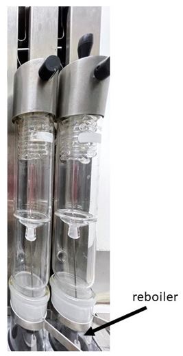
Figure 1: Photograph of Reboiler
FIGURE 2
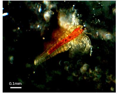
Figure 2: Photograph of Artemia salina used for Toxicity Tests
FIGURE 3
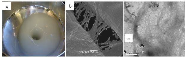
Figure 3: Photographs of Nanofibrils of Peach Palm: (a) - suspension gel; (b) - SEM Photograph; (c) - TEM micrograph
FIGURE 4
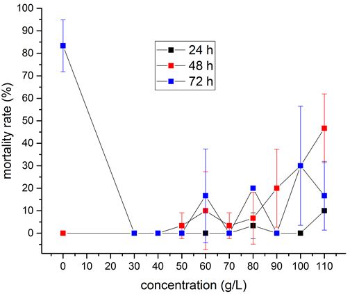
Figure 4: Plots of Artemia salina mortality (in %) Vs. Concentrations of gel with nanofibrils in the saline solution showing toxicity of nanofibrils after 24, 48 and 72 h. The continuous lines are just for guidance. The standard deviations are shown for each measurement
Tables at a glance
Figures at a glance