Synthesis and in vitro Evaluation of A Green Nanomedicine for the Treatment of Patients with COVID-19 Virus and Lung and Breast Cancers
Received Date: March 02, 2022 Accepted Date: April 02, 2022 Published Date: April 04, 2022
doi: 10.17303/jnsm.2022.8.102
Citation: Zahra Fakhroueian (2022) Synthesis and in vitro Evaluation of A Green Nanomedicine for the Treatment of Patients with COVID-19 Virus and Lung and Breast Cancers. J Nanotech Smart Mater 8: 1-21.
Abstract
The Covid-19 pandemic (coronavirus disease 2019) was first identified in Wuhan, China in December 2019 and it has continued to this day in all countries of the world. In this very detailed and novel research, an intelligent and active nanomedicine has been successfully synthesized and formulated for the treatment of coronavirus patients based on green liquid solvent nanofluids. In this important work, green new nano oxidizing and nano reducing agents have been synthesized through co-precipitation-alkali hydrolysis and surface modification methods and used in the formulation of nanodrug. First, the in vitro cytotoxic effects of nanoproduct on human breast cancer cell line (MCF-7) and human lung cancer cell line (A549) were investigated and the IC50 values were determined as 0.1 and 1 ng/ml for MCF-7 and A549, respectively at 50 μg/ml- 0.01pg/ml concentrations range of it. On the other hand, the effect of nanodrug increased and the highest effects were observed after 72 hours. In a second experiment, in vitro tests were performed on standardized SARS-COV2 virus to evaluate the efficacy of nanodrug on the Vero cell line after 24-72 hours. The in vitro results in CCID50 tests showed that a concentration of 0.001 pg/ml of the nanodrug could reduce the number of cultured Attenuated SARS-CoV2 virus by two folds. According to the results obtained, this research should be continued and with the achieved effective dose on the proper animal model to evaluate the systemic toxicity and effect on SARS-COV2 virus under in vivo conditions. The essential characterizations of this nanodrug have been fully performed and zeta potential was measured negative. Totally, the safety dose of this nanoproduct on normal cell lines can have dual valuable properties both for the treatment of breast and lung cancers and also coronavirus-19 patients.
Keywords: COVID-19; Antiviral nanodrug; Coronavirus patients; Oxidation-Reduction; CCID5 tests; Vero cell; MCF-7 and A549 cell lines; Anticancer; Dual property; Pandemic
Introduction
A dual property or dual-targeted therapy for a special nanomedicine indicates a distinct type of its feature in which two different properties can be seen in an intelligent nanodrug which may offer some advantages. That means in such small-scale surfaces, should provide a series of modification that improves the nanomaterials biological properties. With the help of knowledge of nanotechnology and nanomedicine, we questioned having a dual property against coronavirus (covid-19) and breast and lung cancers in this research. In vitro tests of this new nanodrug showed that it has the ability to fight both types of diseases. Which is both economically advantageous for certain patients and from the point of view of modern pharmacy and the development of synergistic medical technology. Because the code of the important relationship between these two properties can be used scientifically and academically.
Coronaviruses (CoVs), which are the largest group of viruses causing respiratory and gastrointestinal infections, are enveloped, spherical or pleiomorphic viruses, and is known to include Alpha, Beta, Gamma, Delta waves and a new strain of the Omicron coronavirus in South Africa [1]. Coronaviruses (CoVs) have a specific morphology and are composed of enveloping viruses containing a non-segmented positive sense, single stranded ribonucleic acid (RNA) virus known as Betacoronavirus in Coronaviridae.
One of the key highlights of this virus is the story of spike proteins that allow these viruses to penetrate host cells and cause infection (The spike (S) protein of coronaviruses facilitates viral entry into target cells). The essential structural proteins of CoVs genome include the spike protein (S), the envelope protein (E), the membrane protein (M), nucleocapsid protein (N) and RNA, which can attach to angiotensin-converting enzyme 2 (ACE2) and serine protease MPRSS2 receptors [2] receptor in the body through active amino acids in its spike glycoprotein (S) structure (crowned tentacles). Therefore, the S protein plays a crucial role in the first step of infections by CoVs, being the leading mediator of viral entry, followed by attachment of CoVs to the surface of host cell’s receptors [3]. Human immune deficiency (HIV), influenza, respiratory syncytial the parainfluenza viruses, would also be the most significant portals and the primary sites of entry for invasion or generating infection.
As initial solutions of humans against this wild CoVs virus: Arbidol, Lopinavir, Favipiravir, Oseltamivir, Ritonavir plus Oseltamivir, Lopinavir plus Ritonavir, Mesenchymal stem cell treatment, Darunavir plus Cobicistat, Dexamethasone, Methylprednisolone, Hydroxychloroquine sulfate and ChloroAbstract quine phosphate chemical compounds together with Remdesivir (Veklury) have been used to treat patients with severe CoVs symptoms when a clinical trial is unavailable or not feasible [4]. However, gradually the power of nanotechnology should enter to help, treating the patients through protection, prevention, diagnosis, treatment and vaccination [5]. Nanomaterials, particularly surface functionalized nanoparticles (NPs) and active quantum dots (Q-Dots), are able to create various innovative methods for mitigation strategies against the effects of CoVs. Gaining experience or gathering experiments, both can conduct us to various metallic and non-metallic NPs such as magnetic, ZnO, CuO, Ag, NiO, ZrO2, iron oxide, CuI and Au/SiO2 NPs, inorganic and metallic-based nanocarriers, and polymeric NPs, as highly promising solution to inactivate the CoVs and covid-19 infection [6]. For example, nanocarriers for the co-encapsulation of many candidate drugs such as Remdesivir and Hydroxychloroquine, or lipid nanoparticle encapsulated mRNA encoding for perfusion of stabilized spike glycoproteins which was the strategy of Pfizer- BioNTech and Moderna companies in their vaccines. Regarding the virus testing and detection, several NPs such as gold (Au) NPs and quantum dots (Q-Dots) NPs, graphene oxide (GO), mesoporous silica, polypeptide, chitosan and carbon nanotubes (CNTs) can also creatively be formulated [7]. Interestingly, in this regard, the authors published a short communication in 2020 about the oxidation-reduction chemical reactions between the CoVs and amino acids of vital cross folding proteins in DNA and RNA in the body, based on chemistry science, for the first time [8]. Cancer is a disease caused by gene mutation, leading to an uncontrolled cell division. Being recognized but poorly understood phenomena. Nowadays, nano-based systems are strategically designed to enhance and increase the therapeutic index of anticancer nanodrugs through improvement of their bioavailability, stability and residency at targeted lung regions [9]. Based on this fact, a new and promising approach for treatment of lung cancer has been developed by researchers at Lund University in 2020. Additionally, it has been found that, among all cancers, lung [10], breast [ 11-14] and prostate [15] carcinoma are the three most common fatal cancers [16] and the majority of the newly discovered cases are related to lung and breast cancer [17]. In this territory, also having the specific knowledge of surface modification of NPs, Q-Dots NPs and hydrophilic nanomaterials, in immunotherapy, plays a significant task in cancer treatments [18, 19], pursuing the main goals of increasing the time period of nanodrugs in circulation and enhance their penetration and accumulation in malignant tumors, increasing reactive oxygen species (ROS) production, sensitizing mitochondrial membrane potential (mitochondrial dysfunction) and inducing both early and late apoptosis in cancer cells. Besides, mitochondrial membrane depolarisation is an early key characteristic of cell death and also could affect respiration and increase ROS generation [20].
The creative idea of CoVs beside cancer. This paper is focused on the high potential of dual property of smart nanocomposites to combat virus especially CoVs and also MCF-7 breast cancer and A549 lung cancer cell lines through the design of a new nanofluid (nanodrug) whose main elements are nano-oxidant and nano-reducing agents, which are prominent adjuvants for fine NPs in this nanoformulation for the first time. In anticancer studies, IC50 values were determined as 0.1 and 1 ng /ml for MCF-7 and A549 cancer cell lines respectively, via MTT and LDH assay. And in in vitro anti-CoVs studies against standardized SARS-CoV2 virus and molecular diagnostic tests using standard Real-Time PCR assay was performed. Here, we evaluated the efficacy of nanodrug on the Vero cell after 24-72 hours. The results of antiviral activity revealed that 0.001 pg/ml of the nanodrug could reduce the number of cultured attenuated SARS-CoV2 virus by two folds. Most likely, this dual nature of our nanomedicine could stimulate new big thoughts between virus and cancer cell lines. This nanomedicine is currently being tested for in vivo on animal hamster models.
Materials and Methods
Synthesis and formulation of prominent COVID-19 nanodrug
In the synthesis and formulation of this nanodrug, which is introduced for the first time, the chemical theory of redox-reaction has been used. Therefore, the ideal nanomaterials related to this theory were synthesized by sol-gel, co-precipitation technique and wet chemical methods via alkali hydrolysis and then formulated in suitable and reliable eco-friendly liquids such as wetting agent ethoxylated, pharmaceutical solvent, PEG-polymer as binding agent, biodegradable surfactant based on a natural compounds and sorbitan hydrophilic emulsifier at optimal pH=7-8 conditions. Nano-oxidizing material such as fine zinc oxide NPs and its novel surface modification which has listed as a safe material by FDA and hinder the viruses’ replication in cell culture were synthezied along with nano-reducing agent nanocomposites in this healthy nanodrug. However, no toxic solvents or dangerous compounds have been used in this new formulation for the treatment of cancer and coronavirus disease. The nanodrug is in the form of nanoemulsion, grayish color, dissolves well in distilled water, is very stable and does not precipitate (no agglomeration). The formulation of this nanodrug is based on oxidizing and reducing nanocomposites and synthesized fine nanomaterials through advanced fabrication and chemical modification, where the size of nanocomposites is 20-21 nm and ZnO nanoparticle’s size is between 5-15 nm. The ratio of nano- oxidants to nano-reducing compounds is 2 to 1, which does not harm other vital proteins in the body. Of course, the average crystallite size of dried original nanodrug according to Scherrer equation in XRD pattern was calculated 16.38 nm. Therefore, with these average suitable nanometer sizes, shape, charge, or surface chemistry they can easily enter into chemical reactions with viral nanometer proteins. All the starting materials for the synthesis of the nanodrug have been purchased from the local companies in Iran such as Kimiagran Emrooz, Neutrino, Arsha and Pishgaman Nanomaterials. The final price of the drug is also reasonable. This nanodrug is currently in the animal testing phase.
Part One - Cancer Research:
Cell lines and cell culture studies
The human breast cancer cell line (MCF-7) and human lung cancer cell line (A549) were obtained from the Pasteur Institute of Tehran, Iran. The cell lines were cultured in RPMI-1640 medium supplemented with 10% heat inactivated fetal bovine serum (Gibco), 100 U/ml of penicillin, and 100 μg/ml of streptomycin and incubated at 37 ºC in the presence of 5% CO2 and 95% humid air after 24 h. The cells were harvested at 70–100% confluence with trypsin/EDTA and used for the in vitro cytotoxicity assays.
Determination of the appropriate concentrations of nanodrug for the study of screening experiments
In [10], a multi-sensor fusion technique is proposed. In this case, after the event detection step, appliance features gathered from various domestic devices are fused before applying various unsupervised ML techniques. Multi-sensor fusion aims to reduce probable sensing mistakes by connecting data collected by energy consumption submeters, light, and audio sensors.
In this experiment, 10 different doses of nanodrug were used for two cancer cells during two stages. In the first stage, 200,000 MCF-7 and 150,000 A549 cells were cultured in each plate well of 24 houses. The cells were incubated for 48 hours for proliferation. Doses of 0.1, 1, 10, 25 and 50 μg/ml of the nanodrug were then applied twice. During drug treatment, high concentrations of this nanodrug interfered with the culture system and morphological examination of cells. Furthermore, after 24 hours of incubation at 37 °C and 5% CO2, the dark color of the nanodrug interfered with 3-(4,5-dimethylthiazol-2-yl)-2,5-diphenyl tetrazolium bromide (MTT) cytotoxicity assay and LDH tests. The results were recorded using an Enzyme-linked immunosorbent assay (ELISA)-reader instrument after performing the MTT test on cells treated with different doses. The culture plate of cancer cells with concentrations of 0.1 to 50 μg/ml nanodrug is shown in Figure 1.
In the second stage, doses of 0.1-0.01-0.001 μg/ml and 0.1-0.01 ng/ml were used and 24, 48 and 72 hours after drug treatment, the culture medium was collected for the measurement of LDH. At the same time, the microscopic images of each well were prepared to study the morphological changes of the cells. Microscopic images show that there are no morphological changes at 24 and 48 hours.
Evaluation of morphology of cells under the influence of nanodrug in the MCF-7 cancer cells
The images (A-F) in Figure 2 show the effect of different doses of nanodrug on MCF-7 cancer cell line at various concentrations after 72 hours.
Result 1: At 72 hours, a dose of 0.001 μg/ml of the nanodrug showed the greatest effect on the morphology of MCF-7 cell line.
Evaluation of morphology of cells under the influence of nanodrug in the A549 cancer cells
The images (A-F) in Figure 3 show the effect of different doses of nanodrug on A549 cancer cell line at various concentrations after 72 hours.
Result 2: At 72 hours, a dose of 0.1 μg/ml of the nanodrug showed the greatest effect on the morphology of A549 cell line. This effect has been identified as an increase in cellular apoptosis and the formation of vesicles and apoptotic body in cells.
Investigation of the effect of nanodrugs on the release of lactate dehydrogenase enzyme (LDH)
Lactate dehydrogenase (also called lactic acid dehydrogenase or LDH) is an enzyme found in almost all body tissues. It plays a significant part in cellular respiration, the process by which glucose (sugar) from food is converted into useful and effective energy for body cells.
LDH test was performed on the nanodrug according to the kit instructions (Cytotoxicity Detection Kit (LDH) (Roche) Cat. No. 11644793001) and cytotoxicity % was determined using the following equation.
Cell Viability% was then obtained.
Cell Viability %= (Cytotoxicity%*100)/Positive control
The diagrams of the test (LDH) of nanodrug are shown in Figure 4.
Evaluation of the effect of nanodrug on the lactate dehydrogenase enzyme (LDH) in MCF-7 cell line
The curves corresponding to the cell viability (% of PC) are statistically shown in Figure 4 using different concentrations (μg/ml and ng/ml) of the nanodrug after 24, 48 and 72 h.
Result 3: The synthetic nanodrug studied showed the highest release of lactate dehydrogenase (LDH) at a dose of 0.1 μg/ml in 24 hours. Elevated lactate dehydrogenase levels usually occur after cell death or when body tissue is damaged. In that case, this enzyme is released into the bloodstream. In addition, Figure 4 shows the lowest release of this enzyme at a dose of 0.01 ng/ml for 72 hours. The LDH test helps determine the location of tissue damage. If LDH is high in the blood, it may mean that certain tissues in the body have been damaged by disease or injury.
Evaluation of the effect of nanodrug on the lactate dehydrogenase enzyme (LDH) in A549 cell line
The curves corresponding to the cell viability (% of PC) are statistically shown in Figure 5 using different concentrations (μg/ml and ng/ml) of the nanodrug for A549 cell lines after 24, 48 and 72h.
Result 4: As an important conclusion from this part of the experiment, according to the curves obtained, the nanodrug showed the greatest effect (0.1 μg/ml) on the release of lactate dehydrogenase (LDH) after 72 hours and in a concentration-dependent manner (p < 0.05) on A549 cell lines.
Anti-proliferative effect of the nanodrug on metabolic activity of the human lung cancer cell line (A549) and human breast cancer cell line by MTT assay including 6 different concentrations compared with control after 72 hours
A significant reduction in the cellular metabolic activity of A459 and MCF-7 cell lines by MTT assay due to the use of advanced nanodrug Covid-19 is shown in Figure 6. To investigate cellular metabolic activity, MTT experiment was performed at 48 and 72 hours and as in the case of cell viability, a significant decrease was observed in the treated cells. As shown in Figure 6, the longer the cells are treated with nanoparticles, the slower their growth rate. Moreover, at higher doses, they inhibit cell growth faster, are more inhibited, and lead to the cessation of the cell cycle in cancer cells.
MTT Experimental results showed that the nanodrug could reduce cellular activity in a dose- dependent and time-dependent manner in A459 and MCF-7 after 72 hours.
Result 5: In this part of the study, the formulated nanodrug showed its highest effect at a concentration of 0.1 μg/ml on the metabolic activity of A549 cell line. In addition, in MCF-7 cell line, the greatest effect was observed at a dose of 0.001 μg/ml. Therefore, the nanodrug has a very effective anti-tumor ability. In addition, using this nanodrug, the metabolism of cancer cells can be precisely targeted and damage to the genome or disruption of cellular metabolic processes can be prevented.
Determination of the dose with the highest lethal effect of nanodrug on MCF-7 and A549 cancer cell lines using standard curves (ST)
This curve is used to compare the survival of the groups with the control group. In fact, the toxicity results of each dose are standardized relative to the rate of normal cell death during culture in the untreated group. Therefore, the rate of baseline cell mortality from drug groups deductions and drug-induced deaths is determined net. According to standard curve of cell survival percentage, the studied nanomedicine showed the highest lethality at a concentration of 0.1μg/ml on the A549 human lung cancer cell line. Furthermore, at a concentration of 0.001 μg/ml, this nanodrug has caused the most lethality on the MCF-7 human breast cancer cell line. It was “golden standard” for cancer detection in this research.
In other words, the basal and normal cell mortalities, obtained from the control group, are deducted from nanodrug induced mortality. The remaining result is the net effect of nanodrugs on cell survival.
Result 6: The optimal doses in terms of toxicity and lethality of cancer cells for our formulated nanodrug are as follows: 0.1 μg/ml for human lung cancer (A549) and 0.001 μg/ml for human breast cancer.
Final conclusion of this part of evaluation of cancer therapy:
Doses of 0.1-0.001 μg/ml nanodrug showed a greater lethal effect on the cancer lines tested. The cellular metabolic activity, which represents vital cellular activity, is further reduced at these doses. On the other hand, with increasing time, the effect of nanodrug improved with the greatest efficacy at 72 hours. Based on the results obtained from this work, the nanodrug showed very high biocompatibility with human lung and breast cancer cell lines tested. Moreover, to determine biosafety, it is recommended to use doses lower than the measured values in the future works.
The effect of nanodrug on the release of lactate dehydrogenase (LDH) and cellular metabolic activity (MTT) for low dose in A549 cancer cell line
In continuation of this interesting research, it was decided to test low doses of nanodrug (such as 0.1 ng and 0.001 pg) in order to determine its performance in low doses in the order of micrograms. Figure 8 shows these findings.
Due to the nature and characteristic of synthetic of the nanodrug, which belongs to the group of nanomedicines, its passage through cell membranes at low concentrations may be high. Therefore, in order to determine the exact effective dose in cell culture conditions, in the next step, doses lower than 0.1 ng/ml were also examined. The results of this section showed that the best lethal dose was 0.01 ng/ml. At lower concentrations, the lethal effect of the nanodrug on cancer cells is reduced.
As observed in Figure 8, according to the results of the experiments (MTT and LDH) carried out using low doses of nanodrugs, doses of 0.1 ng/ml to 0.01 pg/ml did not have a significant lethal effect on A549 cells and the vital activity of the A549 cells, which received these doses from the nanodrug were the same as that of the control group. Thus, ng/ml and pg/ml doses were not used in the subsequent tests. To minimize the side effects and toxicity-related inorganic nanoparticles, nanoagents, and nanocomposites their physicochemical properties (surface functionalization) must be modified. Also, the dosage of the applied engineered nanomaterials (ENMs) must be carefully adjusted [21]. Of course, most recent studies on the side effects of derived from nanomedicine are recommended, especially in their interaction with the immune system.
Part Two- Coronavirus Research
Experimental methods for the in vitro tests of COVID-19 virus on nanodrug
In the second experimental part of this study, the authors focused on the use of the nanodrug as a treatment for coronavirus patients, which is another prominent feature of this product. In this stage, cytotoxicity was investigated and the appropriate concentration of nanodrug was then determined to study the in vitro toxicity of the SARS-COV2 virus.
In vitro evaluation of cytotoxicity and determination of proper concentration of formulated smart nanodrug
According to the previous work on the studied nanodrug, the in vitro toxicity of which was measured in different concentrations on two categories of human cancer cells (A549, MCF-7), the lowest non-lethal concentration of 0.1 pg/ml (1 × 10 -13 g/ml) was measured. In order to evaluate the toxicity of nanodrug and determine the appropriate dose as well as the maximum survival time of the cells treated, the cytotoxicity tests were essential. Initially, concentrations of 0.01 μg/ml (1 × 10 -8 g/ ml), 0.1 ng/ ml (1 ×10 -10 g/ml) and 0.1 pg/ml (1 × 10 -13 g/ml) were prepared and diluted with distilled water from the freshly made main product. Then 5 ml of each coded solution were then tested in the virology laboratory to evaluate the performance of the standardized virus (SARS- COV 2). The test conditions were as follows:
1-Location of viral tests: Amirabad Pharmed Virology Laboratory.
2-Test method reference: Based on the modified method of national standard numbers 16676 and 17981 and internal references of the laboratory.
3-Contact time: 24 to 72 hours.
4-Test temperatures: (in degrees Celsius (25±1)).
5-Greenhouse temperature (in degrees Celsius (37 ± 1)).
6-Specifications of viral strain: (Attenuated SARS-COV2 based on manufacturer’s request). 7- Virus Titer/Viruses Used: 6.2 Log CCID50-TCID50/ml
8-Name of cell lines: Vero cell line
The single layer culture of Vero cell was prepared in 96-well plates and to evaluate the maximum survival time of nanodrug-treated cells, the cells were treated with direct concentrations of three nanoproduct concentrations. The results obtained showed that direct concentrations of solutions in less than 24 hours were toxic to Vero cells and the dilution process should be performed again then to determine the appropriate dose of the nanodrug. In the next step, serial dilutions of ten times were prepared from 0.1 pg/ml solution to evaluate the toxicity of the nanodrug. (0.01 pg/ml, 0.001 pg/ml, and 0.1 fg/ml). The dilutions made did not show obvious toxicity to the cell and were therefore selected to investigate the antiviral effect of exposure to the Attenuated SARS-COV2 virus on the Vero cell.
Investigation of antiviral effect in exposure to Attenuated SARS-COV2 virus on Vero cell lines (African green monkey kidney cells, sensitive to a variety of viruses and high virus titers)
To study the antiviral effect of the nanodrug in various routes of administration of a generic drug of the Attenuated SARS-CoV2 virus on the Vero cell, the virus titration method was used by the method (CCID50) before and after exposure to the drug. The basis of this measurement method is the dilute amount of virus, which infects 50% of cell culture.
1-First, serial dilutions were prepared from the main stock of the virus.
2-100 μL of each dilution were added to 3 houses from 96 well plate cells. One row of wells without virus inoculation and one with non-diluted virus inoculation were considered as a control and a virus control, respectively.
3-The prepared microplates were incubated at 37 °C for one hour to allow the virus to be absorbed into the cells, the microplate being shaken every 15 minutes.
4-The microplate wells were then washed twice with phosphate buffered saline (PBS) and 100 μL of the culture medium containing the nanodrug with the specified concentration were added to each well and transferred to the incubator (the above steps were performed for all three dilutions of the nanodrug).
5-The microplate was checked for cytopathic or cytopathogenic effect (CPE) after 72 hours and the virus titer was determined before and after exposure to nanodrug using the Reed-Manch equation.
Proportionate distance: (% Mortality >50-50) (% Mortality>50 -% Mortality<50) Result report Evaluation of the test results of antiviral activity
Table 1 summarizes the results of the nanodrug antiviral process tests against COVID-19. In this Table, the factors including sample, dilution, contact time, mean virus titer after the test and finally conclusion are observed.
Result of evaluation of antiviral effect on coronavirus:
The results of the antiviral effect test in the presence of SARS-COV-2 virus on Vero cell showed that the dilutions of 0.01 and 0.001 pg of the synthesized nanodrug solution could reduce one logarithm of the corona virus titer. It is noteworthy that in the future animal model tests, we will definitely obtain more realistic results on the living organisms.
Presentation of cytopathic effect for SARS-COV2
In this research, we aimed at detecting the morphological changes induced by coronavirus infection empirically and timely. Figure 9 shows the images of cytopathic effect (CPE)-SARS- COV 2 in comparison to Vero cell and subsequently use virus-specific monoclonal antibody to confirm the presence of virus. Demonstration of cytopathic effects (CPE) induced by virus infection is a practical method (quick identification) to determine the presence of viruses in the clinical specimens [22]. Cytopathic effect must always be based on the comparison with uninoculated cell cultures. In the initial development of cytopathic effects, the SARS-COV2 cells become smaller and rounded as shown in the Figure on the left and cell death induced by severe cytopathic effects and become recognizable under the microscope. In the figure on the right, the nanodrug was able to improve the damaged viral cells by diluting the concentration of 1.100. Our prediction is that by changing this concentration, the percentage of improved cells will definitely increase. Because we chose the lowest concentration to prevent possible toxicity of nanodrug.
Final result from antiviral effect test in the presence of SARS-COV 2 virus on Vero cell The in vitro results in CCID50 test showed that 0.001 pg/ml concentration of the synthetic nanodrug can reduce the number of cultured viruses of Attenuated SARS –COV 2 by ten times. According to the results obtained, it is suggested that the effective dose determined on the appropriate animal model be evaluated for the systematic evaluation of the nanodrug in the future works.
Discussion
Characterization techniques of synthezied nanodrug
SEM images of typical small sized nanoparticles and nanodrug product
Scanning electron microscopy (SEM) (Philips, XL30) images indicate a very fine morphology for ZnO nano oxidant NPs (a), and nanodrug sample (b) (Figure 10). The images show nanospherical morphology for the homogeneous NPs similar to the spherical shape of coronaviruses.
UV-Vis absorption and photoluminescence (PL) spectroscopy of very fine nano oxidant NPs in the nanodrug formulation
The ultraviolet-visible (UV-Vis) absorption spectrum (Perkin Elmer model lambda 35, Perkin Elmer Inc., Shelton, CT, USA) of small ZnO NPs in water based nanodrug (Figure 11 (a)) shows a strong sharp peak at 193 nm (blue emission), indicating that these NPs are very fine and have high band gap energies arising from the strong electronic structures of the defect centers, inner shell electron transitions and finally quantum confinement effects [23,24]. These small NPs with an identical hexagonal wurtzite P63mc crystal structure are able to diffuse into the cell membrane and pave the way for the rest of the nanofluid components to enter the body to fight off the guest coronavirus NPs. In other words, these NPs are at the forefront of this struggle. The room temperature photoluminescence (Varian model carry Eclipse) spectrum of the NPs shows many trapping states and several splitting PL emission wavelengths in the range of 300-510 nm under excitation wavelength of 259 nm (Figure 11(b)) [24].
Importantly, among the new approaches, one of the important similarities between NPs and coronavirus (CoV) is their sizes (mostly reported to be in the range of 70–90 nm), which promotes NPs @ CoV interactions. The closer these dimensions are to each other, the stronger and more stable the connections become. Therefore, SARS-CoV-2 is considered a colloidal particle and a nanoparticle and thus it carries a zeta potential. It is a great point, which the size of the oxidizing agent (electron acceptor) nanocomposite is about 20 nm and the size of the reducing (electron donor) nanocomposite is about 21 nm. Therefore, these drug nanocomposites can easily cross the cell wall barrier of viral proteins (Spike-S) and react biological processes through physicochemically with viral glycoproteins [8].
Determination of Zeta Potential (ζ) for NPs in the form of highly stable nanofluid
Zeta potential measurement is a technique for determining the surface charge of NPs in a colloidal nanosolution. Therefore, ζ potential is a key indicator of the stability of colloidal or nanofluid. In fact, it is an electrokinetic potential in colloidal dispersions based on DVLO theory and determines the adsorption and stability of the particle in any given dispersant medium. In this context, zeta potential (ζ) of one of the fine nano-oxidant NPs in nanofluid was measured as + 50 mV, which indicates a high stability of the synthesized NPs, but the zeta potential of nanodrug was negative. The ζ potential of cells and viruses are mostly amphoteric (negative or positive). It is well known that viruses usually adhere to charged surfaces and charge-charge interaction is often used to remove viruses. In this study, the zeta potential of nano-oxidant and nano-reductant reagents in nanodrug formulation showed negative. But, the spike proteins of many viruses (including influenza and coronaviruses) are positively charged. In this case, between the positive charge of the virus protein surface and the negative charge of the modified surface nanomaterials, strong electrostatic attractions can be generated and the adsorption power between them greatly increases. Then a new complex chemical structure may be formed (in other words, the virus components would be trapped and inactivated). Now, they are able to prevent the virus from entering the body’s host cells (which is also the process of prevention). Since the nanoparticles’ blocking system is a chemical reaction, it is quite clear that the role of pH factor, zeta potential, size and shape of the synthesized NPs should be very important. Why? because if the pH of nanofluid would be acidic, it easily adheres to receptor ACE2. While, if it would be alkaline, it sticks to the Spike S virus protein. Therefore, the pH of the nanodrug solution should be neutral and adjusted between 7-7.5.
Analysis and interpretation of the in vitro properties and performance of coronavirus nanodrug
The World Health Organization (WHO) refers to the coronavirus as SARS-CoV-2 [25] with typical sizes ranging from 60 to 120 nm in diameter. Nanoparticles (NPs) and viruses operate on the same scale, which makes the nanotechnology approach to develop vaccines and safety engineering (immunoengineering) very strong. SARS-CoV-2 can enter into human host cells, which it is key step of its life cycle. Thus, blocking this stage is also critical for prevention of virus infection. Therefore, the mission and task of the nanodrug should be to prevent the virus from nesting in the organs of the body (especially the mouth, throat and lung). However, the best path for the entry of the virus into the body is one through which it can easily arrive and settle in the host’s lungs and gradually disrupt its immune system. The result of researches indicate that ACE2 is a common receptor for SARS-CoV-2 infection in human, monkey, pig and civet cells. The receptor binding domain (RBD) in the SARS-CoV-2 spike can bind directly to the human ACE2 receptor, which can then attach with the spike S@ACE2 through electrostatic potential, electric field lines, electrostatic forces, electrostatic surfaces, and hydrogen bond formation [26,27]. Thus, this binding causes the effective spread of SARS-CoV-2 in large human populations. Besides, as reported, the spike S protein @ ACE2 interaction is an easy target for nanodrugs or vaccines. In fact, they are able to generate protein-protein interaction and new complex structure of SARS- CoV-2 S protein and ACE2 (Spike-ACE2 protein complex) [27]. Actually, blocking this protein- protein interaction inhibits viral entry into the host cell and stops the viral infection without damaging the host cell. Importantly, the function of the nanodrug is to destroy this interaction by neutralizing antibodies and small molecules. Nanomaterials and nanodrugs can be considered as perfect candidates against viral infections; especially COVID-19, because of their potency (size dependence, low toxicity, electrical charge, optical and electronic properties and powerful diffusion) to enter cell membranes easily and interact with viruses and cancerous cell lines and avoid viral genome replication. In this topical new research, the antiviral and anticancer efficacy and mechanism of special NPs, which were applied in this phenomenon, have been highlighted. In particular, it has been attempted to use various specific methods (surface modification) and new mechanisms (redox- reaction) in modern chemistry to create new antiviral functional compounds including nano-oxidant and nano-reductant packages for the first time. It has been proven that virus infectious process generally consists of four steps including: attachment, penetration, replication and budding and the important goal of antiviral NPs or nanodrugs is to operate smartly to limit viruses or block their activity or suppress some or one of these steps [27]. Some of the functional synthetized nanomaterials can directly interact with viruses and cancer cell lines derived from them by physical and chemical reactions and change their capsid protein structures via breaking and dissociation of the susceptible chemical covalent bonds and producing new chains and connections [13]. Overall, if NPs or nanocomposites can intelligently inhibit virus connections, the host cells of the body will be free of infection. So, nanodrugs must have biocompatibility and targeting specific virus. Therefore, it is essential to preserve the antiviral activity of NPs when binding with viruses. NPs must be soluble in distilled water and the solutions must be stable. Currently, drug resistant virus strains are also a critical issue in the synthesis of modern nanodrugs and new functional NPs [28, 29]. The authors of this work were able to determine the function of NPs and how they are adsorbed and permeate into the inner layers of disulfide and thiol bonds (S-S and -SH, respectively) into vital DNA and RNA proteins of the body, while obtaining experimental results on HIV-1 and AIDS. In this regard, the important chemical proposed reactions between the virus, the host cells and cell membranes were determined as oxidation-reduction chemical reactions [8] along with other suggested mechanisms related to bacteria and cancer cells [12-14].
Proposed mechanism of viral cell entry of coronavirus to the human body from a chemical perspective
A key to curbing the spread of SARS CoV-2 and the resultant pandemic is to achieve a clear understanding the mechanism of its entry into the cell. Some scientists believe that the surface S glycoprotein of the virus mediates this process by binding to human ACE2 as the entry receptor and producing ACE2@SARS-CoV-2 RBD complex. However, others suggest that spike glycoprotein (S protein) with α-helical coiled-coil structure can cleave to the S1 and S2 domains during viral packaging [30]. The N-terminal domain, referred to as S1, and a C-terminal S2 are responsible for binding to the receptor and fusion, respectively [31]. Many hydrophilic interactions including formation of salt bridges and hydrogen bonding, which are all chemical reactions, take place on the interface of SARS-CoV-2 RBD and human ACE2. Therefore, from the chemical perspective, we can think deeply about the reactions between the virus and the host cell. Virus surface chemistry, primarily the hydrophobicity and surface charge of the virus, determines its mobility and governs its colloidal behavior in virus adsorption processes. Viruses are well known to adhere to charged surfaces and charge-charge interaction is often used to remove viruses. In this way, there is limited information on the surface chemistry of virus. When coronaviruses contact the surface of the host cells, the spike RBD binds the tips of one lobe to begin the viral entry [32]. Indeed, the virus can make better contact with the N-terminal helix of ACE2 structure and have higher affinity. Generally, infection is initiated by the interaction of the viral particle (nm) with the specific vital proteins on the target cell surface. Coronaviruses are able to exploit many cell surface molecules such as proteins and carbohydrates in body to obtain entry into the target cells [33]. During the course of this research, it was found that two main groups including oxidizing NPs and strong reducing NPs must be present in the formulation of a nanodrug made for patients with breast and lung cancers and COVID-19 virus. Because in reaction and attack to the coronavirus, both of these significant nano groups are needed. The presence of two different groups in nanodrug with different antiviral mechanisms causes a synergistic or advanced therapeutic effect. The authors describe the process of their proposed mechanism as follows:
* The structure of coronavirus is enveloping (Lipoprotein and lipopolysaccharide) and penetrates the host cell through the spike-S protein.
* From a chemical point of view, amino acids make up the structure of proteins. Protein structure is the linear sequence of amino acids and disulfide bonds, which can be ionized as ionic bond or salt bridge (-CH2-NH3+ - O-C=O) in a favorable media and due to the pair of electrons on nitrogen and oxygen are able to form strong hydrogen bonds (-CH2-OH…...O=C-CH2) and disulfide bridges or bonds or disulfide linkage (-CH2-S-S-CH2-) and finally hydrophobic interactions or dispersion forces (CH3-CH-CH3), located in the polypeptide backbone in protein structure. The disulfide bond is highly non-polar (hydrophobic) and there are two ways for breaking them; namely, reduction and oxidation reactions [8]. In fact, disulfide bridges are the cross-linking portions of a polypeptide. They can bind –SH radicals through oxidation reactions to – S-S- covalent bonds in proteins (coupling of two thiol groups). However, being about 40% weaker than C−C and C−H bonds, the disulfide bond is often the “weak link” in many molecules and is subject to cleavage by polar reagents.
* Once the virus enters the cytoplasm of the host cell, under the influence of intracellular enzymes, the enveloping protein of the virus breaks down and the nucleic acid (polynucleotide) is released
From now on, the nucleic acid (biopolymer) of the virus becomes the governor of the infected cell and controls the cell’s protein making apparatus for its proliferation and various mutations.
* At this point, the virus can be considered a living unit. In other words, the nucleic acids (RNA and DNA) of a virus are living units.
* The virus looks at a macromolecule of vital proteins in the human body, which are large polymers of amino acid agents and peptide bonds linked together by a variety of chemical bonds. Penetration through these macromolecules requires a lot of energy (heat, light, proper pH, hydrolysis reactions, ROS production in mitochondria, oxidative stress and etc.), which is beyond its control. In this significant process, the virus can covalently attack the disulfide bonds in the body’s vital protein structure. These bonds hold the polypeptide structure together like a strong base. Access to other links (R-group and hydrophobic interactions, ionic, polar, and hydrogen bonds, and van der Waals interactions) and their agents is a complex task, which that is not possible for the virus.
* With the genetic mutations and various structural changes constantly observed in the corona virus, it appears that it can exchange electrons; that is, it can give or receive electrons. Because, when the structure of a virus changes, the bonds are displaced and therefore the forces needed to form or break the links are created.
* Importantly, without the spike protein, viruses like the novel SARS-CoV-2 would not be able to interact with the cells of potential hosts such as animals and humans to cause infection. In addition, protein S is a critical component in the reaction to NPs and could be an ideal target for vaccine and antiviral research.
* The coronavirus can break the disulfide bond through its spike S tentacles and produce two groups of thiols by a reduction reaction. R-S-S-R (Cystine) + ne‒ to 2R-SH (Cysteine).
* The NPs in the nanodrug have two main responsibilities against these bond disorders.
* First, the nano-oxidant moves toward the thiol groups and takes the free electrons and finally oxidizes them. By doing this, the broken disulfide bonds are regenerated and the immune system is restored. In this chemical process, the NPs themselves are reduced and release electrons. Therefore, NPs must be able to move and exchange electrons. Just like the structure of the coro navirus, which is able to change the its structure and produce a variety of genetic mutations. In this regard, a study done by Abo-zeid et al showed that iron oxide NPs, both Fe2O3 and Fe3O4, can interact with viral spike S glycoprotein destroying its ability of host cell attachment.
Interestingly, they could produce reactive oxygen specious (ROS) that inactivates SARS-CoV-2 at interface of NPs and Spike-S surfaces [34].
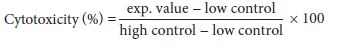
* The second important task of the nanomedicine is very interesting at this stage. The strong nano- reductant containing high permeability, can penetrate into the virus spike S protein and reduce the disulfide bonds. Interestingly, the nanoreducing agent can produce the same disruption previously caused by the coronavirus in the vital proteins of the human body (so-called mirror action) [8, 35]. In this regard, the application of nanomedicines with their specific structural characteristics and physicochemical properties can be a promising therapeutic approach to win the battle between CoVs and host cells [36-38]. Diagram 12 summarizes the topic and mechanism of action of the new nanodrug in the treatment of breast and lung cancer cell lines as well as in the treatment of COVID-19 disease. These communications have been interpreted from a chemical-nano biotechnology perspective [39].
Conclusions
The COVID-19 pandemic represents a global crisis and no effective therapy has yet been proven for it. This study highlights the potential contribution of a new nanomedicine against the recent SARS-CoV-2. This nanodrug is very stable in distilled water and green solution at neutral pH. The essential components of nanodrug are nano- oxidant and nano- reductant agents in selective healthy solvents. First, toxicity of the nanodrug has been investigated on human lung cancer (A549) and human breast cancer (MCF-7) two cancer cell lines in vitro. The nanodrug was evaluated by screening (LDH) and (MTT) cell viability tests using different concentrations of the it (microgram, nanogram and picogram per ml) at 24, 48 and 72 hours. The IC50 values were determined as 0.1 and 1 ng/ml for MCF-7 and A549, respectively. Moreover, the effect of nanodrug continually increased and the highest effects were observed after 72 hours. In the second part of this interesting research, the in vitro evaluation of nanodrug on standardized SARS-COV- 2 virus was performed on the Vero cell line in the range of 24-72 hours. The results of CCID50 experiments showed that using a concentration of 0.001 pg/ml of the nanodrug could reduce the number of cultured Attenuated SARS-CoV2 viruses by two folds. The results of our research indicated the dual properties for new nanodrug formulation. It is encouraging to continue working with this active nanodrug specially to perform animal model tests in reasonable doses at certain times. Therefore, it may be used for prevention, treatment, detection and finally fighting against COVID-19, breast and lung cancers and new mutated variants of coronavirus. Author’ proposed mechanism is based on oxidation-reduction chemical reactions and the production of a nano coating or nano barrier that prevents viral cells from entering the receptor (ACE2) through hydrogen bond formation with amino acids in the ACE2 receptor. Our valuable and benevolence goal of this national research is to save the lives of clinical patients and reduce outbreak of viral diseases.
Acknowledgments
The authors would like to thank the distinguished professors: Reza Zarghami, Mohammad Hassan Panjehshahi, Behzad Rostami, Ali Vatani, Siavash Riahi, and Omid Tavakoli, for their fruitful, continuous and promising supports in approving and advancing the COVID-19 project in the University of Tehran, Iran. Similarly, a sincere gratitude is due to Mrs. Elahe Ahmadi, a skilled graphic designer from the Institute of Petroleum Engineering, University of Tehran, for the graphical support and a very unique thanks to the Arena Diagnostics, Ltd. Iran for their authentic knowledge on COVID-19. In addition, the authors of the paper are very grateful for the valuable efforts of Dr. Mohammad Reza Lornejad to perform nanomedicine on human cancer cell lines in vitro experiments (Pioneer Research Anahita).
Conflict of interest statement
The authors declare that the research was conducted in the absence of any commercial or financial relationships, which could be construed as a potential conflict of interest. In addition, the authors declare no potential conflicts of interest and are responsible for the content and writing of the manuscript.
- Pal, M., Berhanu, G., Desalegn, C., Kandi, V. Severe Acute Respiratory Syndrome Coronavirus-2 (SARS-CoV-2): An Update. Cureus 2020, 12(3): e7423. doi: 10.7759/cureus.7423
- Varahachalam, S. P., Lahooti, B., Chamaneh, M., Bagchi, S., Chhibber, T., Morris, K., Bolanos, J. F., Kim, N.Y., Kaushik, A. Nanomedicine for the SARS-CoV-2: State-of-the-Art and Future Prospects. International Journal of Nanomedicine 2021, 16, 539–560.
- Wrapp, D., Wang, N., Corbett K. S., Goldsmith, J. A., Hsieh, C. L., Abiona, O., Graham, B. S., McLellan, J. S. Cryo-EM structure of the 2019-nCoV spike in the prefusion conformation. Science 2020, 367, 1260–3.
- FDA Emergency Use Authorization: Emergency Use Authorization (EUA) Information, and List of All Current EUAs. Available online: https://www.fda.gov/emergency-preparedness-and- response/mcm-legalregulatory-https://www.fda.gov/emergency-preparedness-and- response/mcm-legalregulatory- and-policy-framework/emergency-use-authorization (accessed on 27 July 2020).
- Tavakol, S., Zahmatkeshan, M., Mohammadinejad, R., Mehrzadi, S., Joghataei, M. T., Alavijeh, M. S., Seifalian, A. The role of nanotechnology in current COVID-19 outbreak. Heliyon 2021, 7, e06841. https://doi.org/10.1016/j.heliyon.2021.e06841
- Wankar, J. N., Chaturvedi, V. K., Bohara, C., Singh, M. P., Bohara, R. A. Role of Nanomedicine in Management and Prevention of COVID-19 Front. Nanotechnol. 2020. https://doi.org/10.3389/fnano.2020.589541
- Nasrollahzadeh, M., Sajjadi, M., Soufi, G. J., Iravani, S., Varma, R. S. Nanomaterials and Nanotechnology-Associated Innovations against Viral Infections with a Focus on Coronaviruses. Nanomaterials 2020, 10, 1072. doi:10.3390/nano10061072.
- Fakhroueian, Z., Esmaeilzadeh, Pe., Esmaeilzadeh, Po. The Mirror Strategy of Nanoparticles Against the Coronavirus. Inter. J. Nanotechnol. in Med. & Engin. 2020, 5 (3), 1-4.
- Rozalen, M., S´anchez-Polo, M., Fern´andez-Perales, M., Widmann, T. J., Rivera-Utrilla, J. Synthesis of controlled-size silver nanoparticles for the administration of methotrexate drug and its activity in colon and lung cancer cells. RSC Adv., 2020, 10, 10646–10660.
- >Crintea, A., Dutu, A. G., Samasca, G., Florian, I. A., Lupan, I., Craciun, A. M. The Nanosystems Involved in Treating Lung Cancer. Life 2021, 11, 682. https://doi.org/10.3390/life11070682
- Jahan, S., Karim, E., Chowdhury, E. H. Nanoparticles Targeting Receptors on Breast Cancer for Efficient Delivery of Chemotherapeutics. Biomedicines 2021, 9, 114. https://doi.org/10.3390/biomedicines9020114.
- Fakhroueian, Z., Dehshiri, A. M., Katouzian, F., Esmaeilzadeh, P. In vitro cytotoxic effects of modified zinc oxide quantum dots on breast cancer cell lines (MCF7), colon cancer cell lines (HT29) and various fungi. J. Nanopart. Res. 2014, 16, 2483. DOI 10.1007/s11051-014-2483- 2.
- Fakhroueian, Z., Esmaeilzadeh, Po., Ghaffari, S. B., Esmaeilzadeh, Pe. Strange Effect of a Synthesized Nanopackage Towards A549, Huh-7, A-375, KB44, MCF-7 And HT29 Human Cancer Therapy. International Journal of Nanotechnology in Medicine & Engineering. 2020, 5(4), 1-19.
- Abdolmohammadi, M. H., Fallahian, F., Fakhroueian, Z., Kamalian, M., Keyhanvar, P., Harsini, F.M., Shafiekhani, A. Application of new ZnO nanoformulation and Ag/Fe/ZnO nanocomposites as water-based nanofluids to consider in vitro cytotoxic effects against MCF-7 breast cancer cells. Artificial Cells, Nanomed. Biotech., 2017, 1-10. DOI: 10.1080/21691401.2017.1290643.
- Ciofani, M. E., Şen, Ö., Çulha, M. Hexagonal Boron Nitride Nanoparticles for Prostate Cancer Treatment. ACS Appl. Nano Mater. 2020, 3, 3, 2364–2372. https://doi.org/10.1021/acsanm.9b02486 .
- >Mao, Y., Liu, X., Bioresponsive Nanomedicine: The Next Step of Deadliest Cancers’ Theranostics. Front. Chem., 2020. https://doi.org/10.3389/fchem.2020.00257 .
- >Zhang, T., Prasad, P., Cai, P., He, C., Shan, D., Rauth, A. M., Wu, X. Y. Dual-targeted hybrid nanoparticles of synergistic drugs for treating lung metastases of triple negative breast cancer in mice. Acta Pharmacologica Sinica. 2017, 38, 835–847.
- Yao, Y., Zhou, Y., Liu, L., Xu, Y., Chen, Q., Wang, Y., Wu, S., Deng, Y., Zhang, J., Shao, A. Nanoparticle-Based Drug Delivery in Cancer Therapy and Its Role in Overcoming Drug Resistance. Front. Mol. Biosci., 20 August 2020 | https://doi.org/10.3389/fmolb.2020.00193.
- Wu, D., Si, M., Xue, H. Y., Wong, H. L. Nanomedicine applications in the treatment of breast cancer: current state of the art. Int J Nanomedicine. 2017; 12: 5879–5892. doi: 10.2147/IJN.S123437.
- Zaidieh, T., Smith, J. R., Ball, K. E., An, Q. ROS as a novel indicator to predict anticancer drug efficacy. BMC Cancer. 2019, 19, 1224, 1-14. https://doi.org/10.1186/s12885-019-6438-y.
- Vahedifard, F., Chakravarthy, K. Nanomedicine for COVID-19: the role of nanotechnology in the treatment and diagnosis of COVID-19. Emergent Mat. 2021, 4, 75–99. https://doi.org/10.1007/s42247-021-00168-8 .
- Wang, T.N., Chao, T. L., Tsai, H. T., Lin, P. H., Tsai, Y. L., Chang, S.Y. Differentiation of Cytopathic Effects (CPE) induced by influenza virus infection using deep Convolutional Neural Networks (CNN). PLOS Comput. Biol. 2020, 16(5), 1-17. https://doi.org/10.1371/journal.pcbi.1007883.
- Ramalingam, V., Varunkumar, K., Ravikumar, V., Rajaram, R. Target delivery of doxorubicin tethered with PVP stabilized gold nanoparticles for effective treatment of lung cancer. Scientific Reports. 2018, 8, 3815 | DOI:10.1038/s41598-018-22172-5.
- Fakhroueian, Z., Katouzian, F., Esmaeilzadeh, P., Bidhendi, S. M., Esmaeilzadeh, Pouriya. Enhanced engineered ZnO nanostructures and their antibacterial activity against urinary, gastrointestinal, respiratory and dermal genital infections. Applied. Nanoscience 2019, 9, 1759– 1773.
- Yang, D. Application of Nanotechnology in the COVID-19 Pandemic. Inter. J. Nanomed. 2021,16, 623–649
- Xie, Y., Karki, C. B., Du, D., Li, H., Wang, J., Sobitan, A., Teng, S., Tang, Q., Li, L. Spike Proteins of SARS-CoV and SARS-CoV-2 Utilize Different Mechanisms to Bind With Human ACE2. Front. Mol. Biosci. 2020. https://doi.org/10.3389/fmolb.2020.591873.
- Verdecchia, P., Cavallini, C., Spanevello, A., Angeli, F. The pivotal link between ACE2 deficiency and SARS-CoV-2 infection. Eur. J Intern. Med. 2020, 76, 14-20. doi: 10.1016/j.ejim.2020.04.037
- Qin, T., Ma, R., Yin, Y., Miao, X., Chen, S., Fan, K., Xi, J., Liu, Q., Gu, Y., Yin, Y., Hu, J., Liu, X., Peng, D., Gao, L. Catalytic inactivation of influenza virus by iron oxide nanozyme, Theranostics 2019, 9, 6920–6935.
- Lara, H. H., Ayala-Nuñez, N.V., Ayala-Nuñez, L., Rodriguez-Padilla, C. Mode of antiviral action of silver nanoparticles against HIV-1, J. Nanobiotechnol. 2010, 8, 1-10.
- Alipoor, S. D., Mortaz, E., Jamaati, H., Tabarsi, P., Bayram, H., Varahram, M., Adcock, I. M. COVID-19: Molecular and Cellular Response. Front. Cell. Infect. Microbiol. 2021, https://doi.org/10.3389/fcimb.2021.563085
- Belouzard, S., Millet, J. K., Licitra, B.N., Whittaker, G. R. Mechanisms of Coronavirus Cell Entry Mediated by the Viral Spike Protein. Viruses 2012, 4(6), 1011–1033. doi: 10.3390/v4061011
- Zhang, Q., Xiang, R., Huo, S., Zhou, Y., Jiang, S., Wang, Q., Yu, F. Molecular mechanism of interaction between SARS-CoV-2 and host cells and interventional therapy. Signal Transduction and Targeted Therapy 2021, 6, 233.
- Nappi, F., Iervolino, A., Singh, S. S. A. COVID-19 Pathogenesis: From Molecular Pathway to Vaccine Administration. Biomedicines. 2021, 9, 903. 1-28. https://doi.org/10.3390/biomedicines9080903 .
- Abo-zeid, Y., Ismail, N.S.M., McLean, G. R., Hamdy, N. M. A molecular docking study repurposes FDA approved iron oxide nanoparticles to treat and control COVID-19 infection. Eur. J. Pharm. Sci. 2020, 153(153), 105465. doi: 10.1016/j.ejps.2020.105465.
- Raha, S., Mallick, R., Basak, S., Duttaroy, A. K. Is copper beneficial for COVID-19 patients? Medical Hypotheses 2020, 142, 109814.
- Abd Ellah, N. H., Gad, S. F., Muhammad, K., Batiha, G. E., Hett, H. F. Nanomedicine as a promising approach for diagnosis, treatment and prophylaxis against COVID-19. Nanomedicine 2020, 15 (21), https://doi.org/10.2217/nnm-2020-0247.
- Alphandéry, E. The Potential of Various Nanotechnologies for Coronavirus Diagnosis/Treatment Highlighted through a Literature Analysis. Bioconjugate Chem. 2020, 31(8), 1873–1882. https://doi.org/10.1021/acs.bioconjchem.0c00287.
- Hamdi, M., Abdel-Bar, H. M., Elmowafy, E., El-khouly, A., Mansour, M., Awad, G. A. S. Investigating the Internalization and COVID-19 Antiviral Computational Analysis of Optimized Nanoscale Zinc Oxide. ACS Omega 2021, 6, 6848−6860. https://dx.doi.org/10.1021/acsomega.0c06046 .
- Yayehrad, A. T., Siraj, E.A., Wondie, G. B., Alemie, A. A., Derseh, M. T., Ambaye, A. S. Could Nanotechnology Help to End the Fight Against COVID-19? Review of Current Findings, Challenges and Future Perspectives. Inter. J. Nanomed. 2021, 16, 5713–5743.
FIGURE
FIGURE 1
Figure 1: Culture plate of A549 and MCF-7 cancer cells with concentrations of o.1 to 50 μg/ml of nanodrug
FIGURE 2
Figure 2: Microscopic images of the effect of different doses of nanodrug on MCF-7 cancer cell line under different conditions of application of doses (A=control, B=0.1, C=0.01, D=0.001 μg/ml), E= 0.1, F=0.01 ng/ml after 72 hours
FIGURE 3
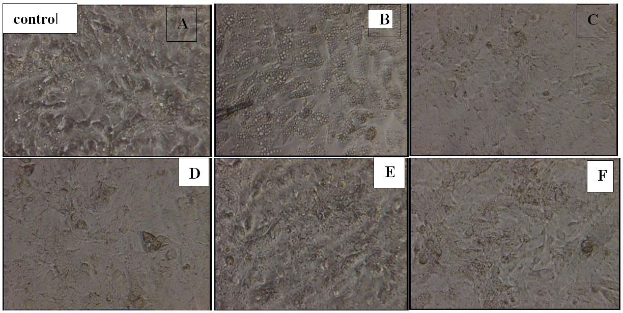
Figure 3: Microscopic images of the effect of different doses of nanodrug on A549 cancer cell line using different doses (A=control, B=0.1, C=0.01, D=0.001 μg/ml), E= 0.1, F=0.01 ng/ml after 72 hours
FIGURE 4
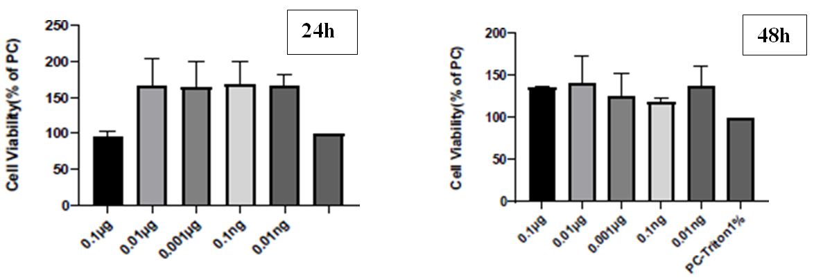
Figure 4: LDH of different types of cell viability factor (PC %)) using various concentrations (μg/ml and ng/ml) of the nanodrug for MCF-7 cell lines in five doses compared to Triton X-100 1% as a control, (# = p<0.05)
FIGURE 5
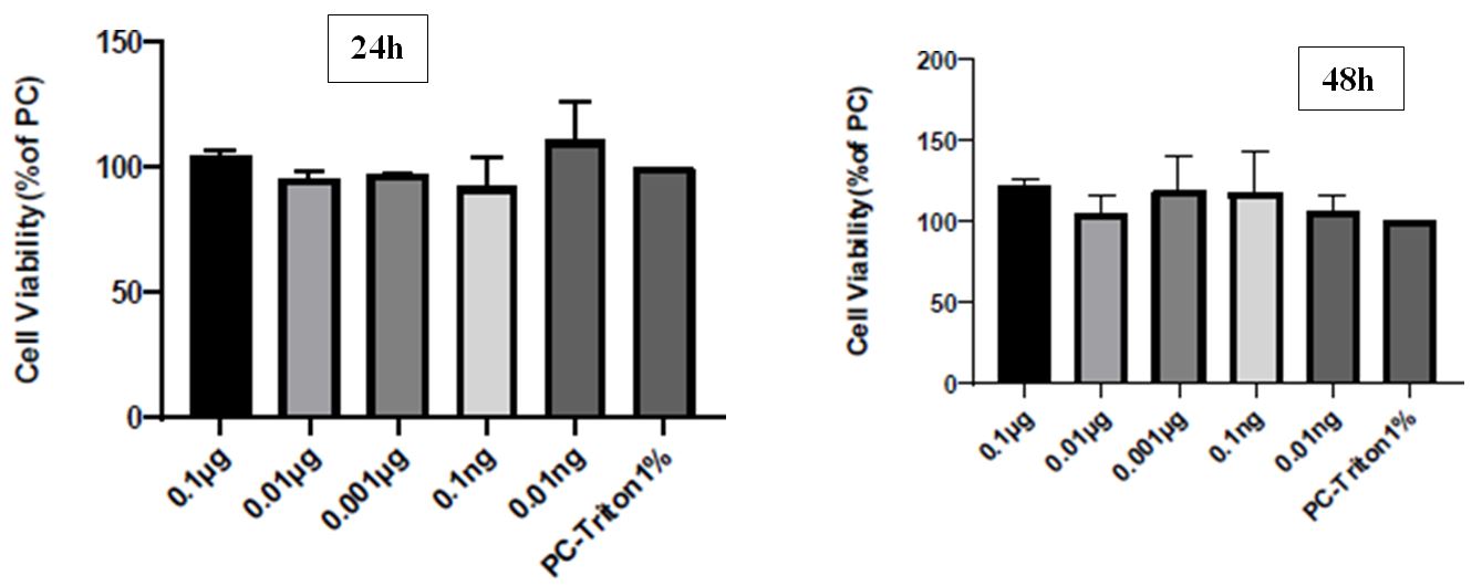
Figure 5: LDH presentation of different types of cell viability factor (PC %)) using various concentrations (μg/ml and ng/ml) of the nanodrug for A549 cell lines, including five doses compared to Triton X-100 1% as a control, (# = p<0.05)
FIGURE 6
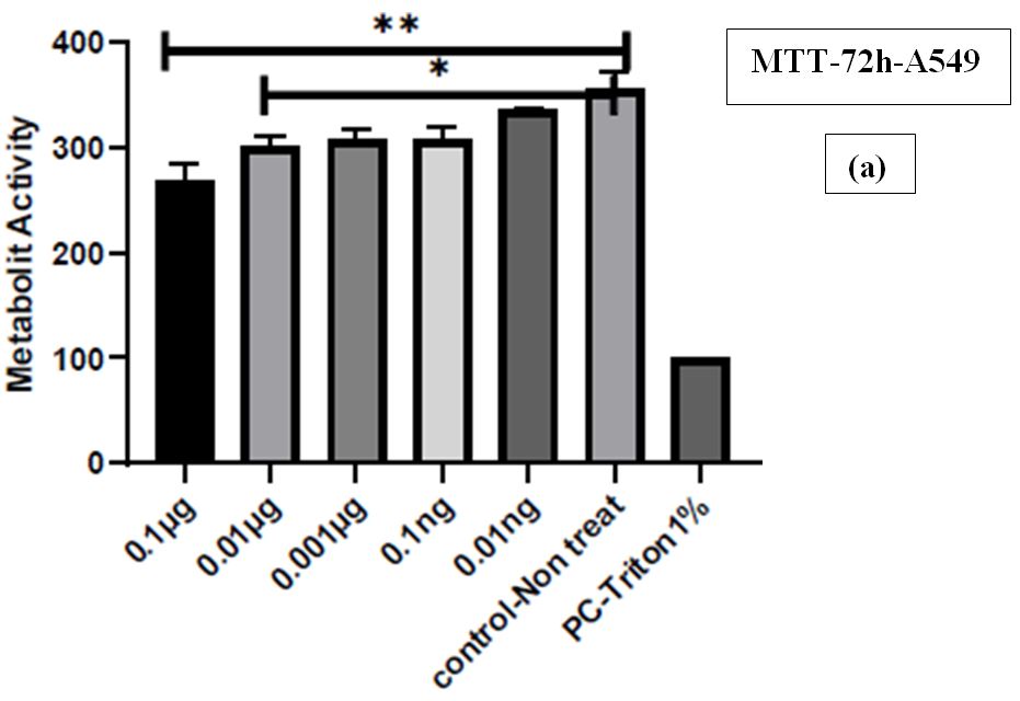
Figure 6: Reduction in cellular metabolic activity of A459 (a) and MCF-7 (b) cell lines by MTT assay using advanced synthetic nanodrug after 72h
FIGURE 7
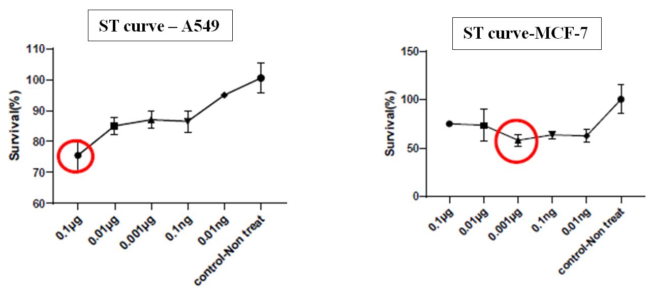
Figure 7: Standard diagram survival % curve in terms of 5 selected doses compared to the control group for A549 and MCF-7 cell lines
FIGURE 8
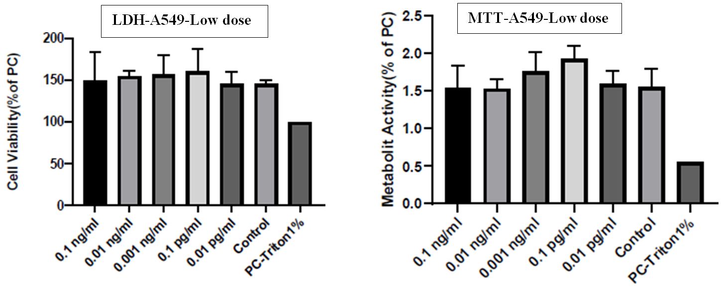
Figure 8: Demonstration of three curves, LDH, MTT and ST at low doses of nanodrug for A549 cell lines. The lowest non-lethal concentration of 0.1 pg/ml was reported
FIGURE 9
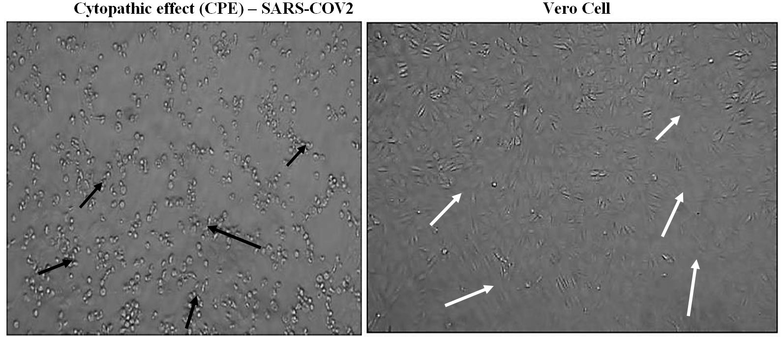
Figure 9: Images of cytopathic effect (CPE)-SARS-COV2 in comparison to Vero cell. This refers to structural or cell morphology changes for a period of time in host cells caused by viral invasion. These pictures may help the diagnosis of viral disease. Black arrows indicate SARS COV 2-induced cytopathic effects and the location of the structure damage and destruction. White arrows show the area where all infected cells were completely detached and floating in the culture medium and are healthy (dose-limiting toxicities in patients)
FIGURE 10
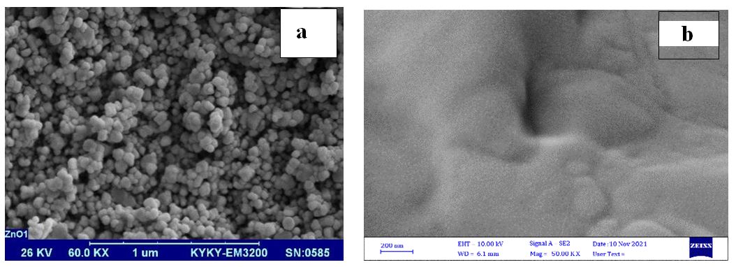
Figure 10: SEM images of the spherical surface morphology of ZnO NPs (a), and nanodrug product solution (b) which contains various types of nanoparticles in nanofluid
FIGURE 11
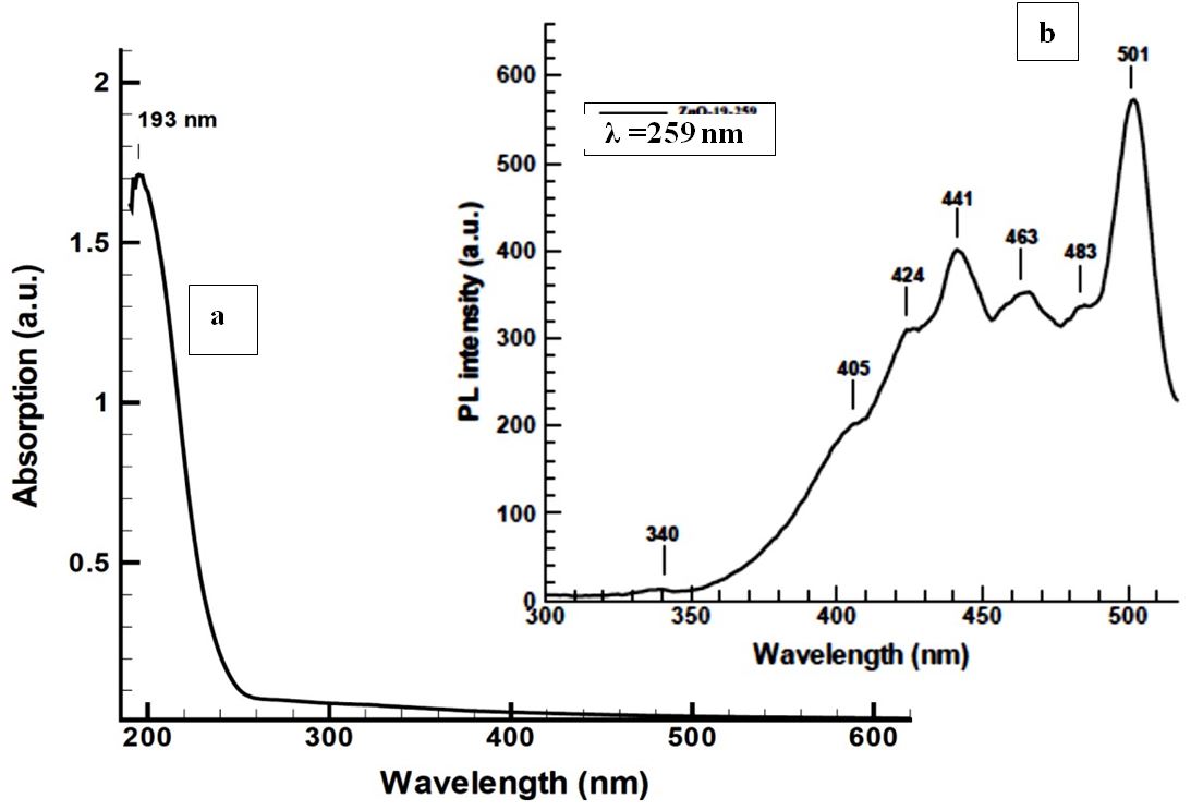
Figure 11: Nanoparticles exhibited high absorption peak at 193 nm in UV-visible (a) and individual photoluminescence emission in the UV-Vis region (b) containing types of surface oxygen vacancies and trapping states as high ladder shaped peaks
FIGURE 12
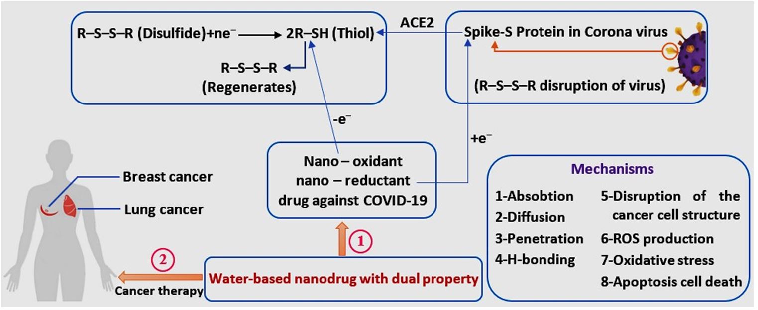
Figure 12: Indicates the dual properties of nanodrug for use in the treatment of COVID-19 and cancer
Tables at a glance
Figures at a glance