
Carbon Fibre Reinforced Polyether-Ether-Ketone (PEEK) Plate Fixation of Open-Wedge High Tibial Osteotomies: Preliminary Results and Complication Rate
Received Date: September 18, 2026 Accepted Date: September 28, 2026 Published Date: October 01, 2026
doi:10.17303/joa.2026.2.101
Citation: Markus Strickner, Michael Ostermann, Alessa Krawinkel, Michael Osti (2026) Carbon Fibre Reinforced Polyether-Ether-Ketone (PEEK) Plate Fixation of Open-Wedge High Tibial Osteotomies: Preliminary Results and Complication Rate. J Osteol and Arthro 2: 1-12
Abstract
Purpose: This study evaluates the radiological and functional results and complication rates following fixation of medial open-wedge high tibial osteotomies (HTO) with a peek-carbon composite stabilizing system (PEEK Power plate).
Methods: 45 patients (mean age 47.7 ± 13.4 years) were included in a retrospective cohort study. Intra-, postoperative and complications to follow-up were recorded and VAS-, KOOS-, Tegner-, Lysholm- and IKDC scores were used to evaluate functional results, anatomical medial proximal tibial angle (aMPTA), anatomical femorotibial angle (aFTA) and anatomical posterior proximal tibial angle (aPPTA) to evaluate pre-, postoperative, implant and follow-up radiographs. Perioperative data were assessed directly during patient admittance; follow-up data were collected at visits to the outpatient clinic and postal questionnaires for at least one year postoperative.
Results: The implant related complication rate was 8.8%. Statistical analysis revealed significant mean improvements in VAS-, KOOS-, Tegner-, Lysholm- and IKDC scores. Postoperative correction of aMPTA and aFTA averaged to 90.7 ± 2.3° and 3.9 ± 2.2° valgus, respectively. The corresponding follow-up values were 89.09 ± 1.6° and 3.2 ± 1.7°, respectively.
Conclusions: The complication rate after HTO using a CR-PEEK plate is comparable to previous reports on other fixation devices. Radiological results are favourable and functional improvements significant.
Keywords: high tibial osteotomy, open wedge, PEEK, complication rate, results
Introduction
Medial open-wedge high tibial osteotomy (HTO) is a well-established and commonly used treatment option in patients with symptomatic unicompartmental medial overload and varus malalignment of the knee [1-5]. Since the invention of the technique, various implants have been developed including short locking and non-locking spacer plates and larger plate fixators without spacer bars [6-11]. Differing complication rates and failure modes of these implants have been described [9,10,12-16]. Angular stability in large plate fixators decreased postoperative loss of correction and complications following HTO [6,14,17,18]. To avoid implant failure and maintain the angle of correction, smaller spacer plates require additional bone grafts [7,13]. A novel carbon fibre reinforced polyether-ether-ketone (CR-PEEK) composite has been developed, yielded favourable biomechanical properties and has therefore been recommended for the use in orthopaedic implants [8,19,20]. Compared to titanium plates, an increased static flexural strength and increased lifetime curves at higher load levels have been reported [18]. In vitro testing of PEEK devices for fracture fixation showed similar characteristics compared to commercially used stabilization systems [21,22]. In addition, CR-PEEK implant simulations predicted reduced stresses at the device-bone interface [19]. Preliminary clinical and radiological results using PEEK implants are rare, but tended towards a promising outcome [14,22]. In a clinical and radiological comparison between PEEK and titanium plate fixators, Cotic et al. [8] noted a higher complication rate after stabilisation of HTO with PEEK implants but no difference in functional outcome between the study groups. Nevertheless, the application of carbon reinforced PEEK for medical technologies has been approved for years in spine surgery. The PEEK composites provide biocompatibility and radiolucency to ensure a clear view onto the fracture or osteotomy site during surgery and on postoperative radiographic imaging [20,23].
The complication rates and the functional and radiological results of CR-PEEK fixation plates for HTO still remain a knowledge gap, having only been investigated by Cotic et al., yet [8,24]. The purpose of this study was to assess the preliminary outcome following open-wedge HTO using the PEEK Power fixation plate (Arthrex Inc., Naples, FL, USA). The hypothesis was that HTO fixation using the CR-PEEK device is associated with similar complication rates and functional and radiological outcome measures comparable to other HTO fixation systems while offering better radiologic assessment options and offering better biocompatiblity
Materials and Methods
Forty-five knees in 45 patients who had undergone HTO with a carbon fibre reinforced polyether-ether-ketone plate (PEEK Power Plate, Arthrex Inc., Naples, FL, USA) for medial unicompartmental osteoarthritis of the knee and concomitant varus malalignment between 2010 and 2012 were evaluated (Fig. 1 and 2). The group included 35 male and 10 female patients with a mean age of 47.7 ± 13.4 years (range, 17.6 to 74.0 years) at the time of surgery. Twenty-three patients (51.1%) were operated for the right and 22 (48.9%) for the left knee. All patients initially presented with symptoms of unicompartimental medial overload originating from varus malalignment of the leg with medial femorotibial osteoarthritis. For preoperative and follow-up assessment of clinical parameters, visual analogue scales (VAS), KOOS-scores, Tegner-activity levels, Lysholm- and subjective IKDC-scores were obtained. Radiological evaluation was conducted using anteroposterior and lateral standard knee radiographs and standing long cassette images of the lower limbs preoperatively, postoperatively, at time of implant removal and at follow-up. In addition, computed tomography (CT) scans of the proximal tibia including the knee joint were obtained on the second day after surgery to evaluate postoperative results (Fig. 3). Radiological measurements included anatomical medial proximal tibia angle (aMPTA), anatomical femurotibial angle (aFTA) and anatomical posterior proximal tibia angle (aPPTA, Fig. 1). Negative values indicate varus deviation, positive values valgus deviation. Complications occurring intra- and postoperatively or in the course to follow-up examination were recorded.
Statistical analysis. Paired t-test for matched variables was used to compare continuous data before and after intervention. To analyse contingency tables the two-tailed Fisher’s exact test was applied. Statistical significance was defined as p<0.05.
The study protocol is in accordance with all legal requirements regarding ethics committee approval.
Results
All fortyfive patients were invited for follow up exams by postal invitation. Patients not responding to follow up invitations for at least 12 months were excluded from the study Twenty-four patients (53.3%) were available for follow-up after 2.1 ± 1.1 years (range, 1.2 to 5.8 years).. Mean body mass index was 27.6 ± 5.0 kg/m2 (range, 16.6 to 38.8 kg/m2). Osteotomy wedge sizes ranged from 3° to 12° (mean 5.7 ± 2.4°). To fill the osteotomy gap, 33 patients (73.3%) underwent autologous cancellous bone grafting. A 3 cm skin incision was performed over the anterior iliac crest and two bone cylinders were harvested. Additional arthroscopic surgery was necessary in 28 patients (62.2%). Table 1 presents additional procedures in detail. Implant removal was performed in 33 cases 0.8 ± 0.3 years after the initial operation (Fig. 2).
Complications
The not implant-related complication rate was calculated with 4.4% (Table 2). On postoperative CT-scans a fracture of the lateral cortex of the tibial head was documented in 11 (24.4%) cases (Fig. 3). Only 6 of them (54.5%) were visible on plain standard radiographs (p>0.2). Additional lag screw fixation was performed in 5 cases (11.1%), 3 of them (6.6%) presented with delayed union which, however, had no adverse effect on final outcome. The implant-related complication rate was 8.8% and arose from 3 delayed unions and one patient (2.2%) who developed non-union and required revision surgery with conversion to a TomoFix plate fixator. All other osteotomies healed uneventful. Specific complications and accordant treatment are demonstrated in Table 2.
Radiological results
Preoperative radiological findings, postoperative angle of correction and corresponding values at time of implant removal and follow-up examination are displayed in Table 3. The preoperative values for aMPTA, aFTA and aPPTA averaged to 85.3 ± 2.1° (range, 79.2 to 88.8°), -2.8 ± 2.3° (range, -0.1 to -9.6°) and 85.3 ± 2.9° (range, 78.5 to 89.7°), respectively. The corresponding values on postoperative radiographs were 90.7 ± 2.3° (range, 86.5 to 97.4°), 3.9 ± 2.2° (range, 13.1 to 0.4°) and 82.5 ± 3.9° (range, 70.2 to 89.1°), respectively, and averaged to 90.1 ± 2.5° (range, 86.2 to 98.5°), 3.4 ± 1.7° (range, 8.0 to 0.8°) and 82.3 ± 4.3° (range, 70.6 to 88.2°) on radiographs after implant removal. Upon follow up evaluation aMPTA, aFTA and aPPTA averaged to 89.9 ± 1.6° (range, 87.1 to 92.8°), 3.2 ± 1.7° (range, 7.0 to 0.0°) and 82.7 ± 4.4° (range, 70.5 to 87.9°), respectively, revealing a loss of correction of 0.7° for aMPTA, of 0.6° for aFTA and of 0.2° for aPPTA, respectively (Table 3).
Functional results
The results for preoperative and follow-up clinical evaluation utilizing VAS-, KOOS-, Tegner, Lysholm- and subjektive IKDC-scores are demonstrated in Table 4. The improvements averaged to 1.5 points for Tegner activity level, to 26.1 points for Lysholm-score, to 24.0 points for IKDC-score and to 3.7 points for VAS, respectively, between preoperative and follow-up values, which represents a statistically significant difference for all scores (p<0.05). Preoperative and follow-up subscales of KOOS are displayed in Table 5.
No statistical significant correlation was detected between BMI (below or above 30 kg/m2), age or gender and overall or implant-related complication rate. Correction angles below or above 7° yielded no statistically significant correlation neither to overall nor to implant-related complications. Statistically significant more fractures of the lateral cortex of the tibial head were detected for an age above 60 years (p<0.03).
Discussion
The results of the present investigation provide functional and radiological data to assess a novel fixation device for HTO regarding effectiveness and safety, which seems to be mandatory for the use of innovations. Several reports in literature detected the insufficiency of novel implants and therefore recommended against their use 10,11. The implant related complication rate in this study was found to be acceptable with only 2.2% of patients requiring revision surgery for malunion. Delayed union occurred in 6.6% without any impact on final functional and radiological outcome. With the use of the PEEK plate, the not implant related complications like infection or haematoma were found to arise less often when compared to other fixation devices 8,16. This confirms the results of Cotic et al. 8 who reported a higher infection rate with the use of larger plate fixators compared to the Peek device. The advantage of a small and slim implant design might not only positively affect painful local soft tissue irritation 4 but also reduce the risk of superficial or deep wound infection (Fig. 2).
Non-union rates following HTO are associated to the type of system used for fixation. Small spacer plates without bone graft yield a higher complication rate especially when used for increased correction angles 5,12,15. On the other hand, the bulky design of plate fixators might negatively influence the risk for infection, which was reported to be as high as 10% 16-25. Valkering et al. 16 found a radiological consolidation process of 10.4 months after Tomofix stabilization of HTO. Non-union rates after HTO with plate fixators range between 0% and 7% in literature 8,16. We did not find risk factors for non-union associated to the use of the presented CR-PEEK plate. The majority of patients underwent cancellous bone grafting. Harvesting two bone cylinders from the anterior iliac crest through a small skin incision was found to be simple and unproblematic and did not result in any patient complaints. Filling the osteotomy gap with bone autografts will certainly also account for the low non-union rate in this series. In a recent study, Cotic et al. 8 directly compared Tomofix and PEEK-plates in a matched-pair analysis and refused from recommending the PEEK plate for clinical practice. Matched pairs were recruited from as database of 160 HTO patients stabilizied with Tomofix plates. The authors found an overall complication rate of 19% and 12%, respectively and a non-union rate of 15% and 0%, respectively. Functional parameters improved significantly in both groups and radiological mesurements revealed a consistent and stabile fixation by both devices. Our investigastion confirms the clinical and radiological results of Cotic et al. 8 and revises the potential complication rate for PEEK implants, which was found to be 2.2% for non-union and 6.6% for delayed union in this series.
Postoperative CT scans facilitated the detection of occult fractures or fissures of the lateral cortex on plain radiographs indicating a more frequent occurrence of a complication potentially endangering osteotomy healing. All cases with delayed union and non-union were associated to a lateral cortex fracture. Nevertheless, no adverse effect on the final functional or radiological outcome could be detected, but a direct positive correlation between uneventful healing in spite of lateral cortex fractures and the use of the PEEK plate can not be drawn from our results. Equally, the use of CT scans as a postoperative routine is not recommended.
The radiological and functional outcome following HTO with PEEK plates is compareable to other fixation devices 3-5,7,8. The CR-PEEK implant is capable to maintain correction angles during osteotomy healing indicating a permanent and stable fixation, which is reported to be a crucial influencing factor in stabilizing osteotomy gaps 6,9,14.
The advantageous material characteristics of CR-PEEK composites provide increased static flexural strength and an increased lifetime curve at higher load levels resulting in favourable biomechanical properties for stable HTO fixation 8. Several authors concluded from histological examinations on acute and chronic tissue inflammation that PEEK plates could be safely used for HTO from a biological point of view 8,22. The advanced concept of multidirectional interlocking screws for plate fixation, the radiolucency of the implant that facilitates intra- and postoperative evaluation of the osteotomy gap and its osseous consolidation as well as anatomic and elaborate shape and design represent further appreciated advantages for a novel HTO fixation device.
Several limitations of this study interfere with an unconfined transfer of our results into clinical practice: we conducted a prospective case series without a control group and evaluated a relatively small sample size with only 45 patients available for baseline assessment and 24 cases for follow-up. Nevertheless we belive in the informative and preliminary quality of our report that might serve as a basis for further research and support surgeons in their decision which implant to choose for HTO fixation. Furthermore, our data confirm the recent findings of Cotic et al. 24, who reported also significant improvements in VAS, WOMAC, and Lysholm scores after 24 months and no loss of correctaion regarding MPTA and PPTA at the time of implant removal in 28 patients following HTO with PEEK plates and an osteotomy gap of 12 mm or less even without bone grafting. The authors found a total complication rate of 4 % caused by one non-union. The non-union rate in this series was 2.2%, all other documented complications did not require revision surgery or were not implant-related.
The complication rate after HTO using a CR-PEEK plate is comparable to previous reports on other fixation devices. Radiological results are favourable and functional improvements significant. Our results indicate that the application of CR-PEEK plates is effective and reliable and can be recommended for HTO fixation.
Funding Declaration
The author(s) received no financial support for the research, authorship, and/or publication of this article
- Aglietti P, Buzzi R, Vena LM, Baldini A, Mondaini A (2003) High tibial valgus osteotomy for medial gonarthrosis: a 10-to 21-year study. J Knee Surg. 16: 21-6.
- Floerkemeier S, Staubli AE, Schroeter S, Goldhahn S, Lobenhoffer P (2013) Outcome after high tibial open-wedge osteotomy: a retrospective evaluation of 533 patients. Knee Surg Sports Traumatol Arthrosc. 21: 170-80.
- Hui C, Salmon LJ, Kok A, Williams HA, Hockers N, van der Tempel WM, Chana R, Pinczewski LA (2011) Long-term survival of high tibial osteotomy for medial compartment osteoarthritis of the knee. Am J Sports Med. 39: 64–70.
- Niemeyer P, Schmal H, Hauschild O, Heyden J, Südkamp NP, Köstler W (2010) Open-wedge osteotomy using an internal plate fixator in patients with medial-compartment gonarthrosis and varus malalignment: 3-year result with regard to preoperative arthroscopic and radiographic findings. Arthroscopy, 26: 1607-16.
- Osti M, Gohm A, Schlick B, Benedetto KP (2015) Complication rate following high tibial open-wedge osteotomy with spacer plates for incipient osteoarthritis of the knee with varus malalignment. Knee Surg Sports Traumatol Arthrosc. 23: 1943-8.
- Agneskirchner JD; Freiling D, Hurschler C, Lobenhoffer P (2006) Primary stability of four different implants for opening wedge high tibial osteotomy. Knee Surg Sports Traumatol Arthrosc. 14: 291-300.
- Asik M, Sen C, Kilic B, Goksan SB, Ciftci F, Taser OF (2006) High tibial osteotomy with Puddu plate for the treatment of varus gonartrosis. Knee Surg Sports Traumatol Arthrosc. 14: 948-54.
- Cotic M, Vogt S, Hinterwimmer S, Feucht MJ, Slotta-Huspenia J, Schuster T, Imhoff AB (2015) A matched-pair comparison of two different locking plates for valgus-producing medial open-wedge high tibial osteotomy: peek-carbon composite plate versus titanium plate. Knee Surg Sports Traumatol Arthrosc. 23: 2032-40.
- Dorsey WO, Miller BS, Tadje JP, Bryant CR (2006) The stability of three commercially available implants used in medial opening wedge high tibial osteotomy. J Knee Surg. 19: 95-8.
- Jung WH, Chun CW, Lee HJ, Ha HJ, Kim JH, Jeong JH (2013) Comparative study of medial opening-wedge high tibial osteotomy using 2 different implants. Arthroscopy. 29: 1063-71.
- Schröter S, Gonser CE, Konstantinidis L, Helwig P, Albrecht D (2011) High complication rate after biplanar open wedge high tibial osteotomy stabilized with a new spacer plate (Position HTO Plate) Without Bone Substitute. Arthroscopy. 27: 644-52.
- Nelissen EM, Langelaan EJ, Nelissen RGHH (2010) Stability of medial opening wedge high tibial osteotomy: a failure analysis. Int Orthop. 34: 217-23.
- Orsel S, Altun M, Bekmezci T, Tonbul M, Yalaman O (2006) Early results of medial opening wedge osteotomy in varus gonarthrosis. Acta Orthop Traumatol Turc. 40: 193-8.
- Pape D, Lorbach O, Schmitz C, Busch LC, Van Giffen N, Seil R, Kohn D (2010) Effect of a biplanar osteotomy on primary stability following high tibial osteotomy: a biomechanical cadaver study. Knee Surg Sports Traumatol Arthrosc. 18: 204-11.
- Spahn G (2003) Complications in high tibial (medial opening wedge) osteotomy. Arch Orthop Trauma Surg. 124: 649-53.
- Valkering KP, van den Bekerom MP, Kappelhoff FM, Albers GH (2009) Complications after tomofix medial opening wedge high tibial osteotomy. J Knee Surg. 22: 218-25.
- Lobenhoffer P, Agneskirchner JD (2003) Improvements in surgical technique of valgus high tibial osteotomy. Knee Surg Sports Traumatol Arthrosc. 11: 132-8.
- Staubli AE, De Simoni C, Babst R, Lobenhoffer P (2003) TomoFix: a new LCP-concept for open wedge osteotomy of the medial proximal tibia - early results in 92 cases. Injury, 34: B55-62.
- Feerick EM, Kennedy J, Mullett H, Fitzpatrick D, McGarry P (2012) Investigation of metallic and carbon fibre PEEK fracture fixation devices for three-part proximal humeral fractures. Med Eng Phys. 15: S1350-4533.
- Rhee PC, Shin AY (2013) The rate of successful four-corner arthrodesis with a locking, dorsal circular polyether-ether-ketone (PEEK-Optima) plate. J Hand Surg Eur. 38: 767-73.
- Kurtz SM, Devine JN (2007) PEEK biomaterials in trauma, orthopedic, and spinal implants. Biomaterials, 28: 4845-69.
- Steinberg EL, Rath E, Shlaifer A, Chechik O, Maman E, Salai M (2013) Carbon fiber reinforced PEEK Optima – a composite material biomechanical properties and wear/debris characteristics of CF-PEEK composites for orthopedic trauma implants. J Mech Behav Biomed Mater. 17: 221-8.
- Tarallo L, Mugnai R, Adani R, Catani F (2013) A new volar plate DiPhos-RM for fixation of distal radius fracture: preliminary report. Tech Hand Up Extrem Surg. 17: 41-5.
- Cotic M, Vogt S, Feucht MJ, Saier T, Minzlaff P, Hinterwimmer S, Imhoff AB (2015) Prospective evaluation of a new plate fixator for valgus-producing medial open-wedge high tibial osteotomy. Knee Surg Sports Traumatol Arthrosc. 23: 3707-16.
- Anastognakos K, Mosser P, Kohn D (2013) Infections after high tibial osteotomy. Knee Surg Sports Traumatol Arthrosc. 21: 161-9.
FIGURE 1
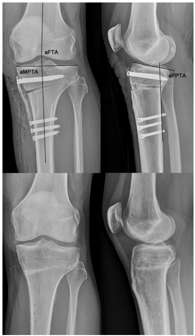
Figure 1: Anterposterior (left row) and lateral (right row) radiographs following HTO with PEEK Power Plate, postoperative (top) and after implant removal (bottom). Measurement method for aMPTA, aFTA and aPPTA.
FIGURE 2
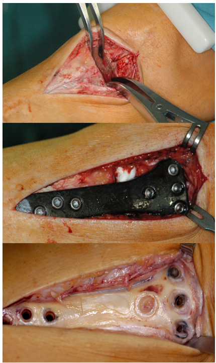
Figure 2: Intraoperative images of a right knee (medial view) demonstrating the osteotomy gap temporariliy expanded with two spreading forceps (top), the PEEK Power Plate in situ and the osteotomy gap filled with corticocancellous graft cylinders from the anterior iliac crest, with absorbable haemostatic gelatin sponges or both (middle) and the osteotomy site status after implant removal (bottom).
FIGURE 3
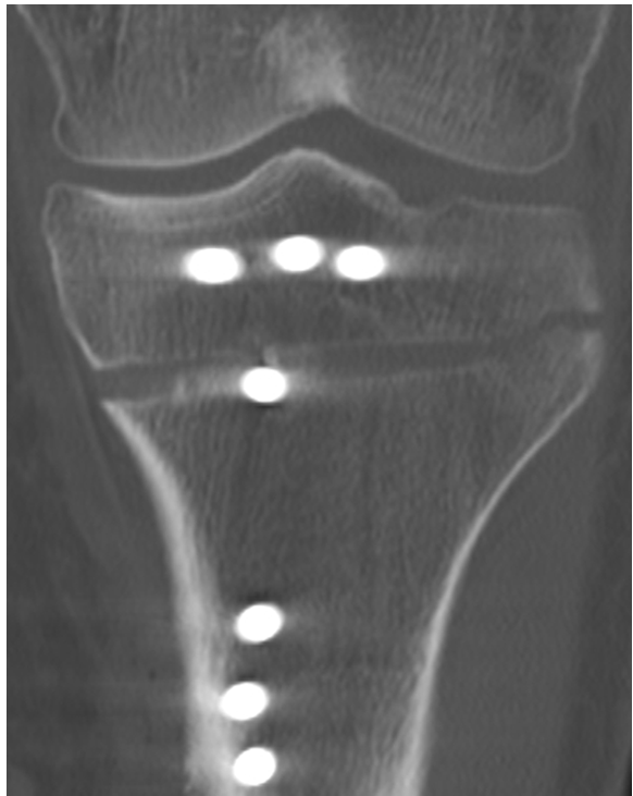
Figure 3: Postoperatice coronar CT reconstruction of a left knee following HTO with fracture of the lateral tibial cortex.
FIGURE 4
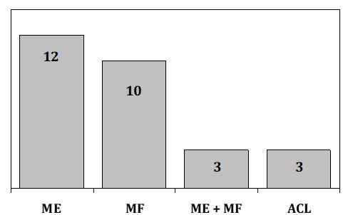
Table 1: Frequency of additional surgery (ME partial medial meniscectomy, MF microfracturing, ME+MF partial medial meniscectomy and microfracturing, ACL anterior cruciate ligament reconstruction)
FIGURE 5
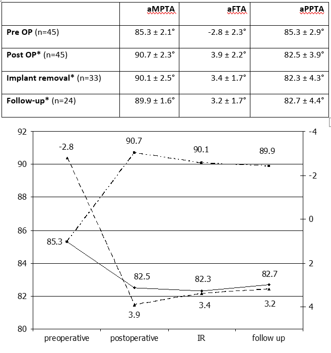
Table 3: Pre-, postoperative, implant removal and follow-up values for aMPTA (■), aFTA (▲) and aPPTA (♦) (Mean ± SD, n.s.not statistically significant between measurements)
FIGURE 6
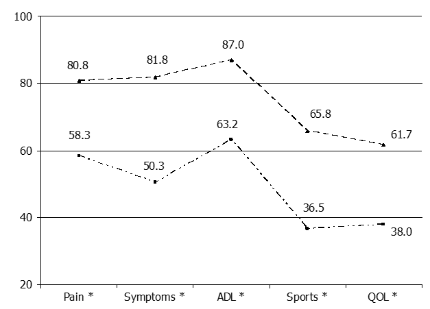
Table 5: Preoperative (■) and follow-up (▲) values for KOOS subscales (ADL activities of daily life, QOL quality of life, * p<0.05 between measurements, n=24)
Tables at a glance
Figures at a glance