Comprehensive Study of Lung Cancer Proton Therapy with Injection of GNPs
Received Date:March 11, 2021 Accepted Date: April 11, 2022 Published Date:April 14, 2022
doi: 10.17303/jocr.2022.3.103
Citation: Reza Khoramdel (2022) Comprehensive Study of Lung Cancer Proton Therapy with Injection of GNPs. JJ Oncol Clin Res 3: 1-22.
Abstract
The use of radiation therapy in lung cancer has shown some exciting and sometimes disappointing advances in recent years.Protons compared with photons interact differently with human tissues, and can be used to improve patient care sufferinglung cancer.A new strategy is the simultaneous injection of nanoparticles with proton radiation to the tumor which has beengiven over a decade to improve conventional radiation therapy. In this work, proton beam therapy (PBT) with GNPs is usedas part of a combination program for the treatment of advanced localized lung cancers. The purpose of this paper is developingthe complex GEANT4 model on the human lung and predict the distribution of absorbed dose in lung tumors during proton therapy without and with high-Z injection of gold nanoparticles. Thus, the distribution of the absorbed dose in lungtumor for four modes of Bethe-Bloch’s relativistic quantum theory, Geant-4 simulation model, Hartree-Fock-Roothaan wavefunctions and the Bortfeld theoretical model without and with the injection of gold nanoparticles in predicted phantom ofthe lung are compared.
Keywords: Cancer; Lung; Nanoparticles; Proton Therapy; Simulation
Introduction
Lung cancer is one of the leading cancers around the world. Lung cancer is categorized into two main categories, depending on the appearance of cancer cells: Non-Small Cell Lung Cancer (NSCLC) and Small Cell Lung Cancer (SCLC). Most importantly, due to the difficulty of treating lung cancer, to a large extent, this cancer can be considered as the most common type of cancer that can lead to death, both in men and in women. It is important to know which type of lung cancer is affected, because small cell cancers have the best response to chemotherapy, while other types, often referred to as large lung cancers, are better treated with surgery or radiotherapy [1].Radiation therapy using photon radiotherapy (XRT) has been a standard treatment for lung cancer since the 1960s [2].Despite years of research, theresult is that treating patients with lung cancer is generally weak due to their cancer tendency to metastasis using XRT. Therefore, studying on a more precise treatment of lung cancer is essential. A potential way to improve the outcome of the disease is optimization radiation therapy. Historically, the patient’s therapeutic outcome is improved if newer radiation patterns are invented. For the first time, radiotherapy was performed through radioisotopes that were directly inserted into the tumor. This was a problem when treating lung cancer from the moment of placing the source in a lung tumor and caused physical damage to the lung. Subsequently, low-energy X-rays were produced that brought KVX-rays into the lung cancer tissue. Unfortunately, these rays penetrated slightly into tissues and deposited the maximum dose to the skin, and deposited only a small fraction of the dose to the tumor. Then, the cobalt isotope with atomic number of 60 was used and gamma rays with two distinct energies of 1.17 and 1.33 million volts (MV) were produced and passed through the skin and resulting in deeper penetration of the dose. Then linear accelerators were developed that produced X-rays or photons with MV energy. These photonic beams penetrated better, but still had deposited a maximum dose between1.5 to 3.5 cm, which gradually decreased the amount of absorption during penetration, so that the radiation without stopping directly was transmitted out of the body. The distribution of X-rays in the body is due to their unique characteristics, which are almost massless and without charge. First, accelerated protons with a large amount of momentum enter to the body, which they can carry a specific depth depending on their initial kinetic energy that is given them by the accelerator (cyclotron or synchrotron). As the proton beam moves to this depth, a relatively small amount of energy is transmitted to the tissue, and gradually, the protons slow down more and more energy is transmitted to the surrounding tissues. The lost energy per unit path length is proportional to the inverse of the square velocity of proton. Shortly before the total proton energy is wasted, the rate of energy loss reaches a sharp peak. When the kinetic energy of the proton is completely deposited in the tissue, the proton inside the body can be rest. The energyof protons in collision with the electrons of neighboring atoms in the surrounding tissues leads to their ionization and thus causes radiation damage. A region in the body where the maximum energy is wasted and the final stop of the protons occurs is narrow at a certain depth depending on the initial energy of the proton beam. This sharpness and peak, which represents the maximum dose due to the charged particles, is called “Bragg Peak”. Beyond this point, as the protons stop, no radiation energy effects on the surrounding tissues .So far, we have just talked about mono energetic proton beams. But the peak of the mono energetic proton beam is very narrow therefore to produce a useful clinical proton beam for the complete coverage of the tumor it is required that in the high-dose area is spread which known as the “spread -out Bragg peak”. Clinical data are necessary to prove the success of the patient’s delivered dose to improve the patient. In the newer proton therapy, uses pencil beam scanning instead of passive scattering and uniform scanning pencil beams are very narrow proton beams that are driven by magnetic fields and move forward and backward in a chess pattern inside the tumor volume. Recently, the use of nanoparticles (NPs) has been suggested as a more effective criterion to improve tumor targeting through radiation effects. When the tumor containing the nanoparticles is exposed under the beam of the proton beam, the antibodies or peptides forming the tumor cells are affected. Therefore, a combination of radiotherapy with nano-medicine opens up a new range of therapies. Hainfeld et al, in 2008 showed that a core of gold nanoparticles with a diameter of 1.9 nm increases the life time of mice exposed to X-rays of 160kV [3].Nanoparticles of gold are already well-known. Other advanced nanoparticles that were made from other heavy elements, such as hafnium [4] and gadolinium [5], which were made by Nanobiotix (Paris, France) and NH TherAguix (Villeurbanne, France), were recently sent to clinics. The effectiveness of high-Z nanoparticles was shown by Kim et al, to improve the proton radiation performance. [6].They injected small nanoparticles (diameters of 1.9 - 14 nm) from gold or iron into tumors of mice that were simultaneously exposed by fast protons with energies of 45 Mev and observed that the absorbed dose in the tumor and, consequently, the killing of cancer cells are increased. The efficiency of gold nanoparticles has been confirmed by Polf et al in increasing the effects of proton radiation through the laboratory [7]. The group observed a significant increase in the mortality of prostate tumor cells with 160 Mev protons when loaded with gold nanoparticles. Recent molecular scale experiments with platinum and gadolinium nanoparticles that have been activated by 150Mev protons [8]. Butterworth and et, al, in 2008, Porcel and et al., in 2010 [10], Jain and et al. in 2014 examined the simulation of three matter suitable treatments such as gold (Au), silver (Ag) and platinum (Pt) such that all of them are compatible with each other and can be used for treatment [11]. Since nanomaterials with highZ can increase the deposited dose in the tumor due to the increase of secondary electrons, we also use simultaneous injection of nanoparticles of three substances (gold, silver and platinum) and proton radiation in the treatment of lung cancer. We will optimize the conditions relative to the state where nanoparticles are not used. For this purpose, in section2 introduces the characteristics of the suggested phantom used in this study, in section2, the Bethe-Bloch model is introduced in a completely relativistic state. In section 3, absorbed dose is described. In the section4, Bethe Block model is given. In sections 5,6 and 7 nuclear and electronic stopping power, Hartree-Fock-Roothaan atomic wave functions and Bragg-Kleemaan’s law are presented, respectively. In the sections 8,9 and 10 multiple Coulomb scattering, proton range and range straggling are described, respectively. In section 11, Bortfeld’s model is presented and finally conclusion is given.
The size of the lung phantom
Phantom of the lung tissue is designed to accurately represent the shape and size of the human lung. The dimensions of the lung phantom are: height 23cm,width 28cm and depth 13.7 (see Figure 1). The GEANT4 toolkit, version 9.2.02, was chosen as the simulation engine. The water phantom with homogeneous geometry and dimensions 30×30×40cm3 was used to simulate GEANT4. Dimension (the beam penetration axis) is determined as the maximum range for proton therapy. The matrix with size of1×1×1mm3 is set to achieve the results of the data. The entire space, with the exception of phantom, is filled with air. At a depth of 10cm inside the phantom, a cubic tumor is placed in a size of . Adipose and skin tissue aredefined in thicknesses of 0.3cm and 0.2cm , respectively.
The phantom material in this geometry is taken from the National Institute of Standards and Technology (NIST). This data provides a detailed mix of materials based on the International Commission on Radiation Units (ICRU). Physical models include electromagnetic and nuclear processes. Electromagnetics physics mainly provides the ionization and multiple scattering of each particle based on Lewis’s theory for the proton. This theory calculates the spatial distribution as the angular distribution after a stage [12]. Low-energy processes, up to 250 electron volts for photons and electrons, like hadrons and ions, are implemented through multiple models. From electromagnetic physics model based on ICRU49 is used for simulation [13]. Nuclear interaction physics is used to study the elastic and inelastic collision between protons and materials. Elastic scattering contributes to the low energy transmission of proton beams while the inelastic scattering contributes to the interaction of nucleon-nucleon along the path of the proton beam. The G4Pre compound model is used for simulation in nuclear physics. This model is suitable for explaining the interaction in the proton beam energy range and is a good estimation for the production of secondary and neutrons particles because it allows the expansion of the low energy range of the hadron kinetic model to inelastic collision of the nucleus- nucleon. For this simulation, the proton beam is used in the energy range 3≤E(MeV)≤ 250MeV with Gaussian distribution σ(E) in the water phantom. The produced Gaussian distribution is compared with the distribution of the simulated and measured dose in the water phantom, respectively. The proton source is a pencil beam scanning type that is located at 10CM far from the upper level of the water phantom. The proton source is parallel to the axis of the beam, (the axis), and 5×10⁶ particles are simulated in our calculations. All particles are examined as if their range exceeds the specified threshold of 0.001mm to be identified. The location of the Bragg peak is the maximum dose point of the curve. The range is defined as the distance between the entrance surface of the beam and the 80% of distal point of the dose in g/cm²
Absorbed Dose
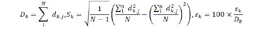
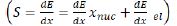
In this paper, we focus on the amount of absorbed dose, which is defined as the deposited energy per unit mass of the matter and is given by: D[GY]=(Edcp[eV]×1.60217646×10⁻¹⁹[J/eV)/(p[kg/cm3]/V[cm3]) Where Edcp is a deposited energy, is the mass density and is the volume of absorbing matter. The absorbed dose Dis a physical quantity and does not reflect the biological effects of radiation. However, D is the first step in assessing the environmental impact of radiation, both for accidental and definitive effects. The Monte Carlo simulation (MC) is known as a required method for the study of nuclear medicine physics, radiology and radiation therapy. The deposited energy and absorbed dose have attracted special interest to radiotherapy programs [14] and imaging programs involving ionizing radiation [15]. In radiotherapy (RT), treatment planning requires an accurate assessment of the distribution of the absorbed dose in the target organ and tissue. Many MC simulators for imaging [16-18] or dosimetry [19-24], have been developed. At present [25], GATE7 [26] is the only MC simulation platform that supports imaging, RT and dosimetry in the same environment. GATE7 is an application GEANT4 toolkit that simulates particles and matter, and GATE7 provides high-level features to facilitate the design of GEANT4- based simulations. The GATE7 is developed by OpenGATE7 and is a community-based initiative that each user can access the source code [27-28] and offer some new features. The GATE7 is essentially useful for a wide range of simulations, including those working on absorbed dose. Despite the fact that GATE7 has been widely accepted and used for a variety of PET and SPECT studies, there are still a limited number of articles that have been used this kind of code in dosimetry. The GATE 7 is provided with a mechanism called Actor Dose that stores the absorbed dose in a specific volume in a 3D matrix. From the point of view of macro- scale GATE7, Actor Dose should be linked to the volume of case study. The user can determine the size of the matrix and the matrix location is defined in the system of coordinated volumetric control. Note that if the user defines the matrix size larger than the associated volume, deposited absorbed dose occurs outside this volume and therefore will not be recorded inside the network.
The Actor calculates the deposited energy (Edep) in MeV and the absorbed dose, D, in Gy. squares of Edep and D are summed and can be used to compute statistical uncertainty when the simulation is split into several branches. Equation (2) defines the statistical uncertainty Σx in pixel , for value, which represents the initial number of events. , is the deposited energy in pixel in the initial event . The absorbed dose can be calculated as the dose in water, which is commonly used in radiotherapy, and the dose in the medium is commonly used in MC simulations [29-30]. This conversion is done by taking into account the relative stopping power and energy transferred through nuclear interactions in a given environment. Note that this conversion method may not be appropriate for some conditions in brachytherapy [31]:
During particle tracking, the deposited energy is added in the matrix for each stage occurring in the connected volume. The two endpoints a step, are a pre and a post points. For a charged particle, a location is randomly selected along the step and values are stored in the matrix at that location. The user must be sure that the length of the step is not very large due to the matrix sampling. The output can be stored in an MHD image format, consisting of a header and a raw data file. The effect of calibration of Hounsfield units (HU) on materials and density has also been reported in reference [32]. At GATE7, the user can manually assign a substance to any HU range, or use Schneider’s method based on a predefined material group (24 materials by default). Two mixtures with the same elemental composition, but different densities are considered as two distinct materials. Each requires the calculation of the cross-section and the stopping power, and if a lot of materials are used, it can be problematic. In reference [33], the authors described a method for dynamically changing the density at run-time, but this technique is still not available in GATE7. In GATE7, the number of materials can be controlled by the tolerance parameter [34]. It is used to decompose a substance into two substances when its HU range is greater than tolerance.
Bethe- Bloch model
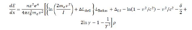
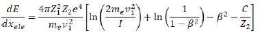
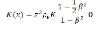
We do not discuss the Bethe-Bloch model in detail, but just examine the aspects of this model for proton therapy. The average energy loss rate per unit length of a relatively relativistic heavy charged particle is described in detail by the Bethe-Bloch equation [35]:
The symbols used in this equation are shown in Table 2, respectively. In a low-energy region (less than 10MeV) , when the particle velocity is equal to one of the target electrons (≈ 0.0073c),ion neutralization due to electron capture plays a crucial role in the stop process, and must be converted to Zeff , which a semi-experimental relation : Zeff=z(1-exp(125zβ-2/3)) has been extracted from Barks experimental data. In Figuresand 3a and b,c,d,e,f,g and h,using the Maple programing , we plotted the three-dimensional variations of different parameters such as β=v/c , Barkas correction, shell correction, Lindhard-Sorensencorrection,[36] density effect, the effective atomic number,the total stopping power and absorbed dose in terms of the energy of the proton beam in the range of 1≤E(MeV)≤250 and the depth of penetration in the lung tissue in the range of 1≤x(cm)≤30 without the injection of nanoparticles of metal. As shown in the diagrams, each parameter plays a specific role in the calculation of the stopping power and absorbed dose, and they are a function of the depth of the proton penetration in the tissue and the energy of the proton.
Nuclear and electronic stopping power
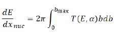
An energetic ion that penetrates the material loses its energy mainly through two processes that are independent of each other. These two processes are: loss of nuclear energy and loss of electron energy. Thus, the stopping power can be divided into two parts: i) nuclear and ii) electronic stopping power. Nuclear stopping power is calculated by integrating over all of the impact parameters:
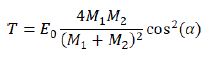
According to Fig.4, the ion with mass of M1 and initial energy E0 is deflected by the target atom with mass of M2. The location of M2 relative M1 to is called the impact parameter, which we represents it by b. During the collision, M1 and M2 deflects with the angles α and β, respectively, relative to the original M2 pathway. During the collision, the kinetic energy, T , it transfers from M1 to M2. According to momentum and energy conversation, this transmitted kinetic energy can be calculated. This kinetic energy T is a function of the angle α , projectile energy,E0 , projectile mass M1 and the mass of target atom M2 in the laboratory system.
The electronic stopping power in this region is given by the Bethe-Bloch equation:
In Fig. 5, we plotted the variations of total mass stopping power
Hartree-Fock-Roothaan atomic wave functions
The Hartree-Fook method is based on the time independent model of the particle. The idea behind this model is to solve the Schrödinger equation for moving electrons in nuclear potentials and the potential of all the other electrons. This repetition continues until the density of the resulting electron charge converges with the electron charge density at start point for one repetition. The Hartree-Fock-Roothaan atomic wave functions are the solutions of HartreeFook’s equation and are represented by the orbital of Slater -type:
In this equation dt is the expansion factor of the orbital.,is the orbital of Slater-type and is characterized by the following equation:
Also, for all modes of without and with the injection of gold nanoparticles, with increasing proton energy, all of the stopping powers are reduced and the minimum stopping power is related to the nuclear interactions. The electronic stopping power is much higher and the main contribution of the total stopping power can be electronic term. Also the total stopping power of without the injection of gold nanoparticles using the model of Hartree-Fock-Roothaan (HFR) atomic wave functions are consistent with the GEANT4 simulation model. In Figure 6 ,the absorbed dose in the lung phantom was compared to with and without the injection of gold nanoparticles in terms of the depth of penetration in the tissue for various proton energy (gold nanoparticles is a sphere with a diameter of 50nm. As shown in Figures 5 and 6, with increasing proton energy, stopping power and absorbed dose decrease, but by increasing the concentration of gold nanoparticles their amount goes up. This is due to the production of secondary electrons. It is seen that from Figures 6a and b,c,and d, with increasing distance and energy, from the beginning of the lung phantom, the absorbed dose at the Bragg peaks is reduced, and the location of the Bragg peak moves with increasing energy to higher x-rays. And the lowest amount of absorbed dose is the related to without injection of gold nanoparticles. By increasing the amount of gold nanoparticles injection from 10 to 75mg/ml, amount of absorbed dose increased by 1.1,1.25,1.45 and 1.75% for 10,25,50 and 75mg/ml respectively in comparison to without injection of gold nanoparticles. This is due to the fact that high-Z nanoparticles, such as nanoparticles of gold, increase the amount of doses deposited inside a tumor or matter due to the increase of secondary electrons and the effect of density. And with an assumption that the tumor is at a depth of 13cm inside the lung with a width of 2cm ,theoptimum energy of the Bragg peak is 93MeV. Our calculations also show that when protons have an energy of 250MeV, they require a phantom with a radius of more than 30cm, so that the protons deposited their energy inside it, and only protons with energy in the range of 80 to 95MeV deposited their energy inside the selected phantom.
Bragg-Kleemaans law
The Bragg-Kleemaan law for the proton range,Ro,is given in terms of the initial energy E and energy curve dE/dx with the following relation:
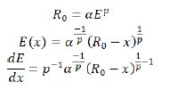
Where α and p , can be determined from the Betheequation or from a model that is fitted with the win-energy data,which their values are: α=0.00262,p=1.736,
In Fig. 7, we plotted the three-dimensional variations of proton mass stopping power and absorbed dose in a lung tissue without the injection of gold nanoparticles using the BraggKleemaan law in terms of proton beam energy in the range of 1≤E(MeV)≤250 and the penetration depth in the range 1≤x(cm)≤30
From the comparison of the graphs for the stopping power of the Bethe-Bloch (Fig. 3-g, using Maple programing) with the model of the Hartree-Fock-Roothaan (HFR) wave functions [37] and the model of proposed phantom of the lung in this work (Fig. 5-C using GEANT4 simulation) without the injection of nanoparticles of gold we found that these models confirm each other.
Multiple Coulomb Scattering
Transitory protons of matter may be deflected by the atomic nucleus, a process commonly referred to scattering, more precisely, multiple coulomb scattering, when observed that angular scattering due to the collective effect of many small single scattering randomly happens. Both protons and nucleus are positively charged particles, as a result, are the interaction between them mainly electrostatics. The multiple Coulomb scattering theory describes the shape of the angular distribution of the particles and its characteristic width. Moliere’s theory gives directly root mean square of multiple coulomb scattering angle at each incident energy for homogeneous slabs of each element with very thin thickness to near stopping thickness.
The mean scatter angleθ0 is determined by the High
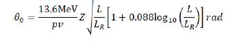
Where p and v are the momentum and velocity of the proton, respectively, is the atomic number of projectile, and L and LR are the target thickness and radiation length, respectively which are the same unit.This formula is valid for a thin slab. The radiation length is the distance which the energy of the radiation particles decreases due to radiation losses as much as the coefficient e^-1(≈0.37) .In Fig. 8, we plotted the variations of the mean Coulomb scattering angle in terms of proton energy in the range of1≤E(MeV)≤250 for with and without the injection of different concentrations of gold nanoparticles in the lung phantom.
As we can see from Fig. 8, with increasing the proton energy, the mean value of the coulomb scattering angle (θ0) increases in all conditions, but the lowestθ0 in the lung tumor is related to the state of the without injection of gold nanoparticles, while with increasing the amount of injection of gold nanoparticles into the lung tumor the value θ0 of gradually increases.
Proton Range
In this work, we use the CSDA method to calculate the proton range. CSDA is calculated by integrating on the initial and final energy of the incident particle on the inverse of the total stopping power, which is given by the following: [37]
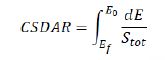
Here,E0 and Ef are the initial and final energy of the charged particle input at the target.In Fig. 9 , we plotted the proton range variations in terms of proton energy in the range of 3≤E(MeV)≤250 without and with the injection of gold nanoparticles in the lung tissue and these diagrams were compared with the results of Hartree-Fock-Roothaan (HFR) [37] wave functions model.
As shown in Fig. 9, the lowest CSDA range is related to the case of without injection gold nanoparticles, and with increasing the amount of the concentration of injection gold nanoparticle, the CSDA has slightly increased such that at of 75mg/mL gold nanoparticles in the lung tissue the CSDA is maximized and in good agreement with results of the HartreeFock-Roothaan (HFR) wave functions model.
Range Straggling
The loss of energy of an ion in matter is a statistical process and it is not definite, and the Bethe equation yields only the energy lost. This change was first described by Bohr, who introduced the concept of energy straggling (σE):
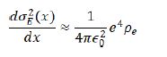
Where is the electron density and is the vacuumpermittivity coefficient. This is valid for energy dissipation thatis large enough to maintain Gaussian approximation. But it issmall enough when ion energy can be assumed to be constant.
In 2004 Schulte and his colleagues gave the following differential equation[39]:
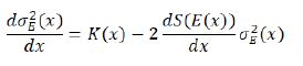
The range straggling (σR) is defined as a function of energy is given by the solution of the following equation:
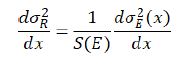
Where S (E) is the total mass stopping power. We calculate the range straggling in terms of proton energy in the lung tissue for two case of without and with the injection of gold nanoparticles, and plot it in Figure 10.
Bortfeld model
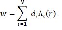
The Bortfeld model is based on the Continuous Slowing Down Approximation (CSDA) with additional features that are not considered in the Bethe Bloch model. In different cases, it is appropriate to have an analytical representation of the Bragg curve instead of using numerically or measured data. The 1997 Brownfield paper provides an approximate analysis of the Bragg curve in a closed form. The validity of this model is based on proton energy between about 10 and 200MeV. The four main principles of this model are: a power-law relation that describes the energy-range dependence, a linear model for reducing flounce due to Columbic nuclear interactions, assuming a fraction of localized released energy, a Gaussian approximation of the distribution of the range straggling and the presentation of the energy spectrum of multi-energy beams with a Gaussian distribution with a linear “tail” [40].From this theoretical model two types of deep doses is achievable:
a) deep dose formula without examining the effects of proton straggling range is [40]:
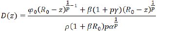
Note that the above equation is valid for z>Ro.For z>Ro,D(z)=0.The above equation can be written as a sum of two terms:
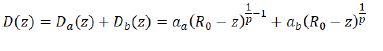
The first term Da(z) Dis the contribution of dose due to proton without nuclear interactions. The second term,Db , represents the dose transmitted by a relatively small fraction of protons having nuclear interactions .
b) Deep dose with the straggling range of the proton’s effects is [40]:
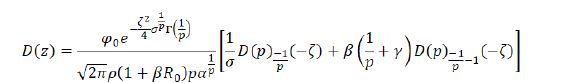
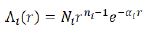
Some of the “special functions” and the parameters that appear in equations (18) to (20) are defined as follows:
Geiger’s law is: Ro=αEoo, the range Rois determinedby the exponent dependence on energy Eo, where and are constant. In ICRU 49,p=1.77 , but the Bragg-Kleeman law describes the constant α .According to Geiger’s law, the proton traverses the maximum range R0 of in initial energy E0. Therefore, in the energy E(x) along x (direction of motion), the proton travels the distance R0-x, which is the same as the solutions of the Weber’s differential equation.Γ(z) is the gamma function defined by Euler’s formula. The functional relationship between and can be given with the following approximation:
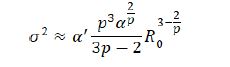
Where α' is a factor which depends on the stopping matter through α.Roand σ are in cm. In the Borteff model, γ is the fraction of energy that is locally absorbed. In this model,γ=0.6 and β≈ 0.012cm-1.
∅
o is the particle flounce that is the number of particles per cm². Suppose .In Fig. 12, the three-dimensional variations of absorbed dose with the efeffects of the proton straggling in terms of the penetration depth and energy of the proton beam in the lung tissue without the injection of gold nanoparticles has been brought.Comparison of Figures 11 and 12 it is seen that in the case of without injection of gold nanoparticles, the absorbed dose value taking into account the effects of the range straggling, is less than the amount of absorbed dose regardless of the absorption effects. Comparing the absorbed dose diagram using completely theoretical quantum relativistic Bethe-Bloch model with the Maple programming in Fig. 3 and the absorbed dose diagram without the injection of gold nanoparticles through the proposed phantom GEANT4 / GATE7 simulation in Fig. 6 and the absorbed dose diagram using Bragg -Kleemaan law with Maple programming in Fig. 7b and absorbed dose diagram using Bertfeld model without considering effects of proton range straggling using Maple programming in Fig. 11 and absorbed dose diagram using Bertfeld considering the effects of proton range straggling using Maple programming in Fig. 12, we find that Bragg’s peak only seen in the Bethe-Bloch model and GEANT4 / GATE7 simulation model of the proposed phantom, and the values of absorbed dose from each purposed model are different, but this difference is not so much, and the proposed GEANT4 / GATE7 simulation model is more complete than the other models, also the results of this proposed model confirm that injection of gold nanoparticles into the lung tumor increases the absorbed dose and the enhancement of concentration of the gold nanoparticles increases the value of absorbed dose.
Conclusion
In this review, we summarize the current status of the proton beam therapy (PBT) for lung cancer. The proton source is a hydrogen nucleus and the proton is accelerated by an accelerator. The proton is stopped at a certain depth, known as the Bragg peak, which significantly reduces the outlet dose and reduces ,damage to healthy tissues around tumor volume. These proton therapeutic properties can be useful in the treatment of lung cancer in patients with cardiovascular disease, with poor pulmonary function or history of previous breast cancer therapy. This work has examined a number of research topics related to proton therapy issues that are important for optimizing the future of cancer treatment. Proton therapy is an important tool in the oncology period, where the radiation therapy goal is to treat a tumor with a minimum toxicity and maximum efficiency. Proton therapy is not expected to be uniform in all scenarios for all patients, but it is expected to dosage be done in such a way that the patient achieves superior clinical results at appropriate times. The proton beams are sensitive to various types of uncertainty, such as respiratory motion, changes in the patient’s location and tumor contraction. But the new proton therapy technology creates a major challenge in radiotherapy planning and delivering. And further research is needed to optimize protons, especially for new beam systems. This requires more understanding of physics to create designs that are strong in the face of uncertainties
- Palma D.A., Senan S., Oberije C., Belderbos J., de Dios N.R., Bradley J.D., Barriger R.B., Moreno-Jimenez M., Kim T.H., Ramella S., et al. Predicting esophagitis after chemoradiation therapy for non-small cell lung cancer: An individual patient data meta-analysis. Int. J. Radiat. Oncol. Biol. Phys. 2013;87:690–696.
- Wolf J, Patno ME, Roswit B, D'Esopo N. Controlled study of survival of patients with clinically inoperable lung cancer treated with radiation therapy. Am J Med 1966; 40: 360-367.
- Hainfeld JF, Dilmanian FA, Slatkin DN, Smilowitz HM.Radiotherapy enhancement with gold nanoparticles. J Pharm Pharmacol 2008; 60(8): 977-85.
- Maggiorella L, Barouch G, Devaux C, et al. Nanoscale radiotherapy with hafnium oxide nanoparticles. Futur Oncol 2012; 80-81, 1167.
- Sancey L, Lux F, Kotb S, et al. The use of theranostic gadolinium-based nanoprobes to improve radiotherapy efficacy. Br J Radiol. 2014;87:20140134.
- Kim J-K, Seo S-J, Kim K-RK-H, et al. Therapeutic application of metallic nanoparticles combined with particleinduced X-ray emission effect. Nanotechnology. 2010;21:425102.
- Polf JC, Bronk LF, Driessen WHP, et al. Enhanced relative biological effectiveness of proton radiotherapy in tumor cells with internalized gold nanoparticles. Appl Phys Lett. 2011;98:193702.
- Schlathölter T, Lacombe S, Eustache P, et al. Improving proton therapy by metal-containing nanoparticles: nanoscale insights. Int J Nanomed. 2016;11:1549.
- Butterworth K T, Wyer J A, Brennan-Fournet M, Latimer C J, Shah M B, Currell F J and Hirst D G Variation of strand break yield for plasmid DNA irradiated with high-Z metal nanoparticles Radiat. Res. 2008; 170 381-7
- Porcel E, Liehn S, Remita H, et al. Platinum nanoparticles: a promising material for future cancer therapy. Nanotechnology. 2010;21:85-103.
- Jain S, Coulter JA, Butterworth KT, et al. Gold nanoparticle cellular uptake, toxicity and radiosensitisation in hypoxic conditions. Radiother Oncol. 2014;110:342–7.
- Lewis H. W., Phys. Rev. 1950;78: 526.
- ICRU . Stopping powers and ranges for protons and α-particles ICRU Report, 1993; 49 (Bethesda, MD)
- Reynaert, N, Vandermarck S, Schaart D, Vanderzee W, Vanvlietvroegindeweij C, Tomsej M, Jansen J, Heijmen B, Coghe M, and Dewagter C. Monte carlo treatment planning for photon and electron beams. Radiat. Phys. Chem.2007; 76: 643-686.
- Murphy MJ, Balter JM, Balter S, BenComo JA, Das IJ, Jiang SB,. Ma GM, Olivera GH, Rodebaugh RF, Ruchala KJ, Shirato H, and Yin FF.The management of imaging dose during image- guided radiotherapy: Report of the AAPM Task Group 75. Med. Phys. 2007;34:4041-4063.
- Ljungberg M and Strand SE.A Monte Carlo program for the simulation of scintillation camera characteristics. Comput Methods Programs Biomed, 1989; 29: 257-272.
- Harrison R and Vannoy S.Preliminary experience with the photon history generator module of a publicdomain simulation system for emission tomography. in Proceedings of the IEEE Nuclear Science Symposium and Medical Imaging Conference ,1993; 1154-1158.
- Cañadas M, Arce P, and RatoMendes P.Validation of a small-animal pet simulation using gamos: A GEANT4based framework. Phys. Med. Biol. 2011;56: 273–288.
- LANL, MCNPX 2.6.0 Users's Guide, Technical Report No. LA-CP-07 1473, 2008.
- Battistoni G, Muraro S, Sala P, Cerutti F, Ferrari G, Roesler S, Fasso A, and Ranft J.The FLUKA code: Description and benchmarking. in Hadronic Shower Simulations Workshop, edited by M. Albrow and R. Raja (AIP Conference, Fermilab,Batavia, IL, 2006; 896,31–49.
- Ferrari A, Sala P, Fasso A, and Ranft J, FLUKA: A Multi-Particle Transport Code, Technical Report No. INFN/TC 05/11, SLAC-R-773, CERN2005-10; 2005.
- J. Perl J. Shin B. Faddegon, and Paganetti H.TOPAS: An innovative proton Monte Carlo platform for research. Med. Phys.2012; 39: 6818–6837 .
- Walters BR, Kawrakow I, and Rogers DW, "History by history statistical estimators in the beam code system," Med. Phys.2002; 29: 2745–2752.
- Kawrakow I and Fippel M. Investigation of variance reduction techniques for Monte Carlo photon dose calculation using XVMC. Phys. Med. Biol. 2000;45: 2163–2183.
- Jan S, Santin G, Strul D, Staelens S, Assié K, Autret D, Avner S ,et al. GATE7: A simulation toolkit for PET and SPECT. Phys. Med. Biol.2004; 49: 4543-4561 .
- Jan S , Benoit D, Becheva E, Carlier T, Cassol F, DescourtP, Frisson T, Grevillot L, et al. GATE7 V6: A major enhancement of the GATE7 simulation platform enabling modelling of CT and radiotherapy. Phys. Med. Biol. 2011;56:881-901 .
- The OpenGATE7 Collaboration, http://www.open-GATE7collaboration.org; 2014.
- The OpenGATE7 Collaboration, http://git.openGATE-7collaboration.org/git/ openGATE7-public.git; 2014.
- Paganetti H, "Dose to water versus dose to medium in proton beam therapy," Phys. Med. Biol. 2009;54: 4399 4421
- Grevillot L, Bertrand D, Dessy F, Freud N, and Sarrut D.GATE7 as a GEANT4-based Monte Carlo platform for the evaluation of proton pencil beam scanning treatment plans. Phys. Med. Biol. 2012;57: 4223-4244 .
- Tedgren A C and Carlsson G A.Specification of absorbed dose to water using model-based dose calculation algorithms for treatment planning in brachytherapy. Phys. Med. Biol.2013; 58: 2561-2579.
- Jiang H, Seco J, and Paganetti H.Effects of Hounsfield number conversion on CT based proton Monte Carlo dose calculations. Med. Phys.2007; 34: 1439-1449.
- Jiang H and Paganetti H.Adaptation of GEANT4 to Monte Carlo dose calculations based on CT data. Med. Phys.2004, 31: 2811-2818.
- Sarrut D and Guigues L.Region-oriented CT image representation for reducing computing time of Monte Carlo simulations.Med. Phys. 2008; 35: 1452-1463.
- Newhauser D and Zhang R. The physics of proton therapy,Phys. Med. Biol.2015; 60: R155–R209.
- Lindhard J and Allan H. So"rensen, PHYSICAL REVIEW A, 1995; 53: 4, 2443-2456
- Metin U , Mustafa C T. Stopping power and range calculations in human tissues by using the Hartree-FockRoothaanwave functions. Radiation Physics and Chemistry ; 2017.
- Highland VL. Some practical remarks on multiple scattering. Nucl Instrum Methods ;1975, 129:497–9.
- Ulmer W and Schaner B. Foundation of an analytical proton beamlet model for inclusion in a general proton dose calculation system. Radiation Physics and Chemistry,2011; 80.
- Bortfeld T. An analytical approximation of the Bragg curve for therapeutic proton beams , Med Phys. ;1997 24,12:2024-33.
TABLE 2
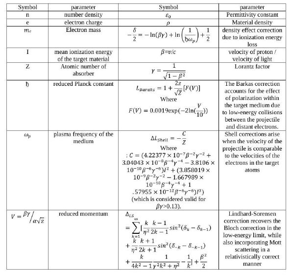

Table 2: List of parameters in the Bethe-Bloch equation
FIGURE 1
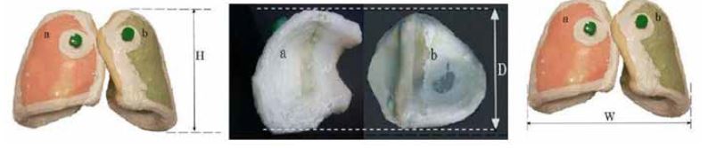
(Hight: H = 23 cm, Width: W = 28 cm and Depth: D = 13.7 cm)
Figure 1: The dimensions of the anatomical lung phantoms a) the right lung b)the left lung.
FIGURE 2
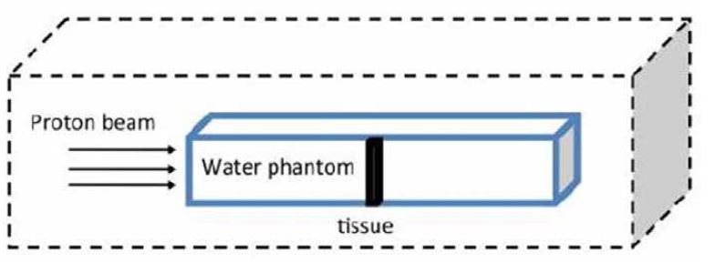
Figure 2: A plan for the selected phantom used in this study
FIGURE 3
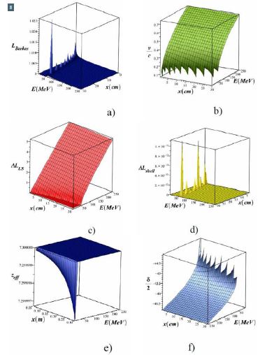
Figure 3: 3D variations of β = v/c, Barkas correction, shell correction, Lindhard-Sorensen correction, density effect, effective atomic number, total stopping power and absorbed dose, in terms of proton beam energy in the range of 1E(MeV)=250 and the penetration depth in the lung tissue in the range of 1=x(cm)=30 without the injection of metal nanoparticles (using Equation 3)
FIGURE 4
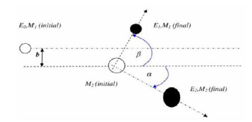
Figure 4: A plan indicating an ion with mass M1 and initial energy E0 that collides to an atom with the mass of M2 and causes it to move with energy E2 and to reduce its E1 energy
FIGURE 5
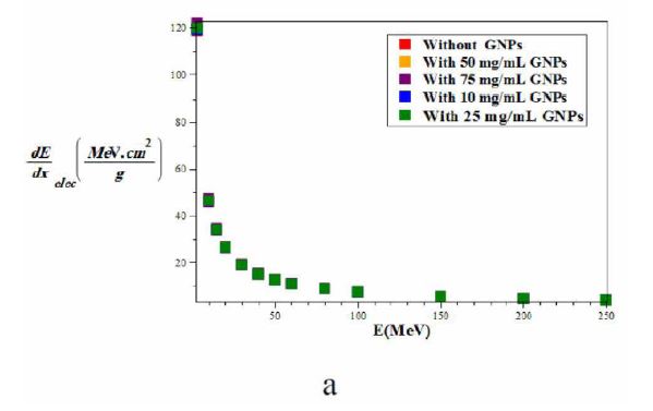
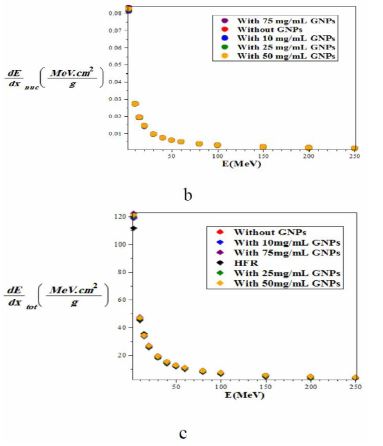
Figure 5: Comparison of the mass-stopping power a) electronic b) nuclear c) total of proton with and without injection of gold nanoparticles with different concentrations in the lung phantom and comparison with the Hartree-Fock-Roothaan (HFR) [37] atomic wave function model without Injection of gold nanoparticles using the GATE7 / GEANT4 simulation From the observation and comparison of the graphs shown in Fig. 5, the minimum of stopping power in all three cases of nuclear, electronic and total, is related to without nanoparticles of gold. By increasing the concentration of gold nanoparticles, the amount of all type of the stopping power gradually and slightly increases
FIGURE 6
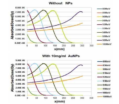
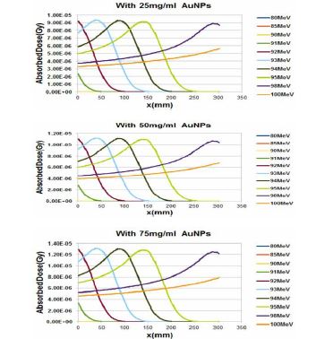
Figure 6: Comparison of the absorbed dose in the lung phantom with or without the injection of gold nanoparticles in terms of penetration depth for various proton energy (gold nanoparticles are considered as a sphere with a diameter of 50 nm and for this calculation we use GATE7 / GEANT4 simulation)
FIGURE 7
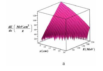
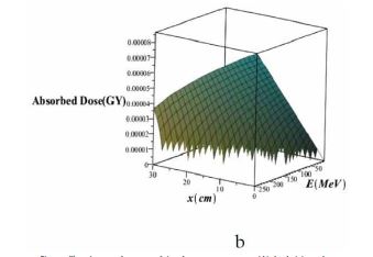
Figure 7: Three-dimensional variations of a) total mass stopping power and b) absorbed dose in lung tissue without injection of gold nanoparticles using Bragg-Kleemaan law in terms of proton energy in the range of 1=E(MeV)=250 And the penetration depth in the lung tissue in the range of 1=x(cm)=30
FIGURE 8
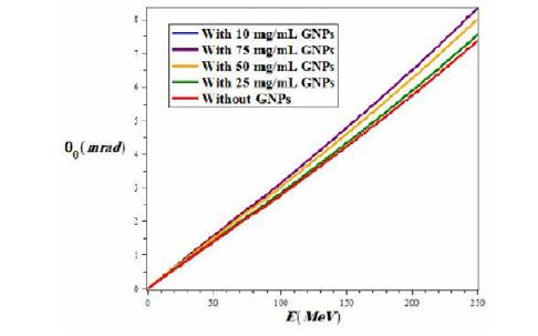
Figure 8: The variations of the mean Coulomb scattering angle in terms of proton energy in the range of 1=E(MeV)=250 for with and without the injection of different concentrations of gold nanoparticles in the lung phantom.
FIGURE 9
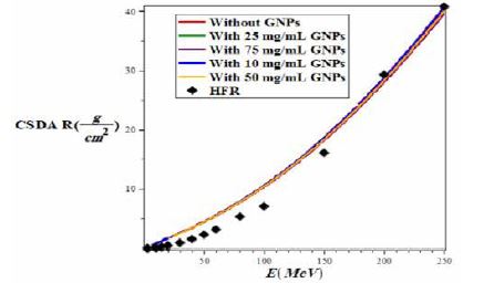
Figure 9: Variations of CSDA proton in lung tissue with and without injection of gold nanoparticles using GEANT4 / GATE7 simulation based on the energy of the proton beam in the range of 1=E(MeV)=250 and the penetration depth in the range of 1=x(cm)=30 and compare it with the model of Hartree-Fock-Roothaan (HFR) wave functions[37]
FIGURE 10
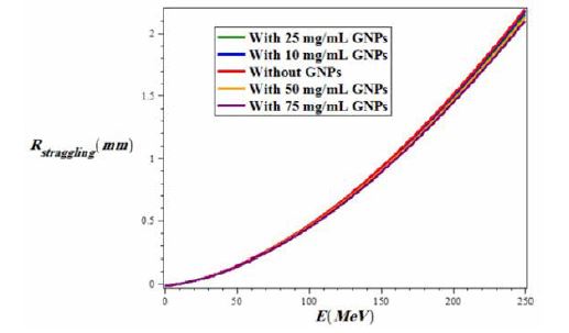
Figure 10: variations of proton range straggling in lung tissue for two states of with and without injection of gold nanoparticles using GEANT4 / GATE7 simulation versus the energy of a proton beam in the range of 1=E(MeV)=250 and the penetration depth in the range of 1=x(cm)=30 As shown in Fig. 10, the range straggling increases with increasing proton energy for both modes of with and without the injection of gold nanoparticles, but the highest range straggling is for the case of without injection of nanoparticles of gold and the lowest amount is related to injection of gold nanoparticles at a concentration of 75mg/ml. In fact, if the amount of injection of gold nanoparticles is more and more, the straggling range decreases.
FIGURE 11
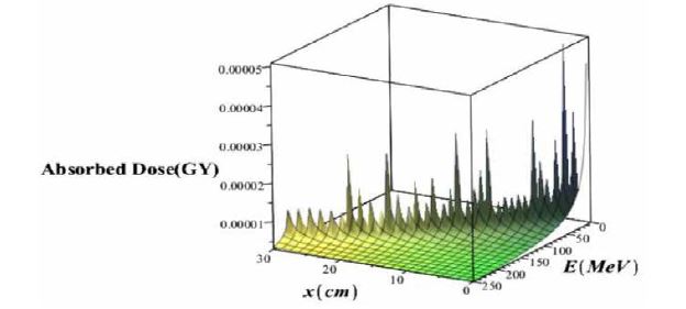
Figure 11: Three-dimensional variations of the deep dose without considering the effects of the proton straggling range in terms of the penetration depth and energy of the proton beam
FIGURE 12
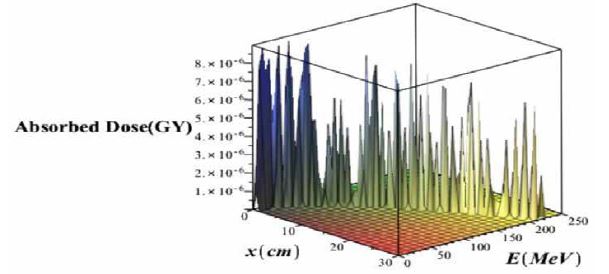
Figure 12: Three Dimensional variations of absorbed dose with considering effects of proton straggling range versus the penetration depth and energy of the proton beam without injection of gold nanoparticles
Tables at a glance
Figures at a glance