
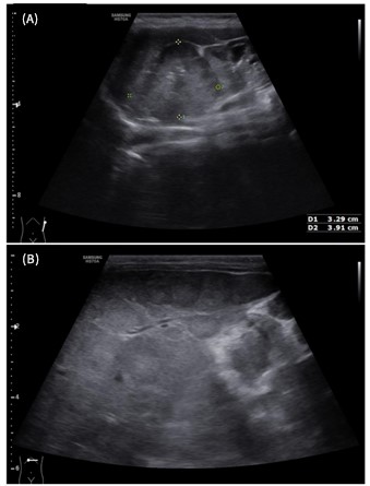
Figure 1: Ultrasound of the left labia majora demonstrating a well-defined, heterogeneous, predominantly hypoechoic solid mass. This sonographic appearance confirmed the lesion’s solid nature of the swelling, prompting subsequent tissue biopsy for definitive diagnosis.
Figures at a glance