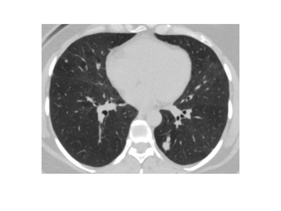
Figure 1: Computed tomography (CT) findings. Computed tomography (CT) examination revealed a 1 cm solid nodule in the left lower lobe
Figure 1: Computed tomography (CT) findings. Computed tomography (CT) examination revealed a 1 cm solid nodule in the left lower lobe
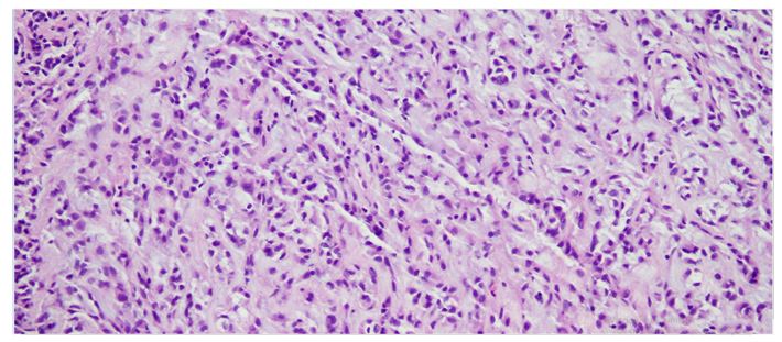
Figure 2: 20X10 HE Endobronchial PPMS. Tumor cells were round and tumor cells are quite small and arranged in a reticular or ribbon-like arrangement in a mucoid background
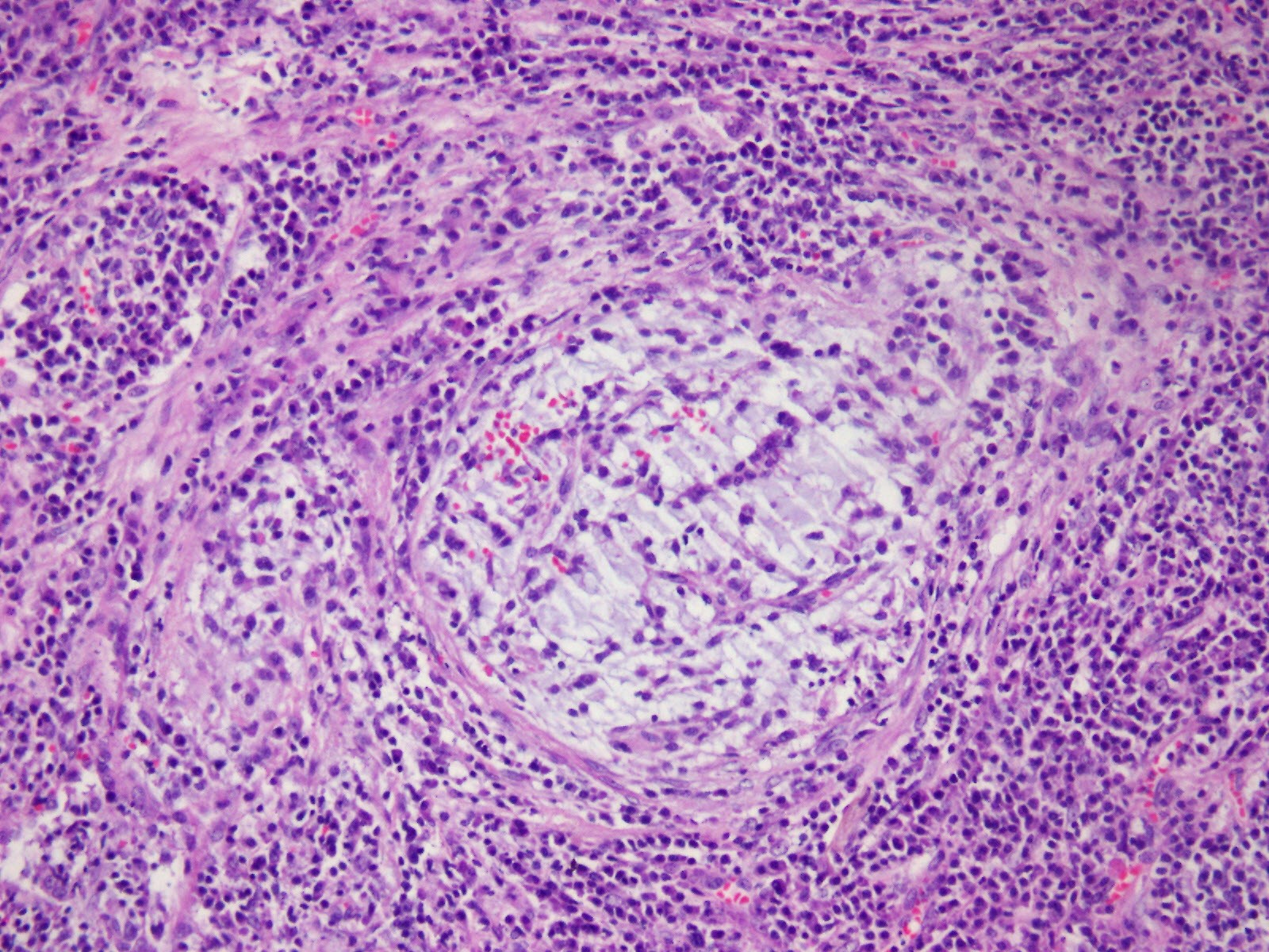
Figure 3: 20X10 HE Tumor cells on myxoid floor, reticular pattern, spindle-shaped, stellate appearance, chronic inflammatory infiltrate
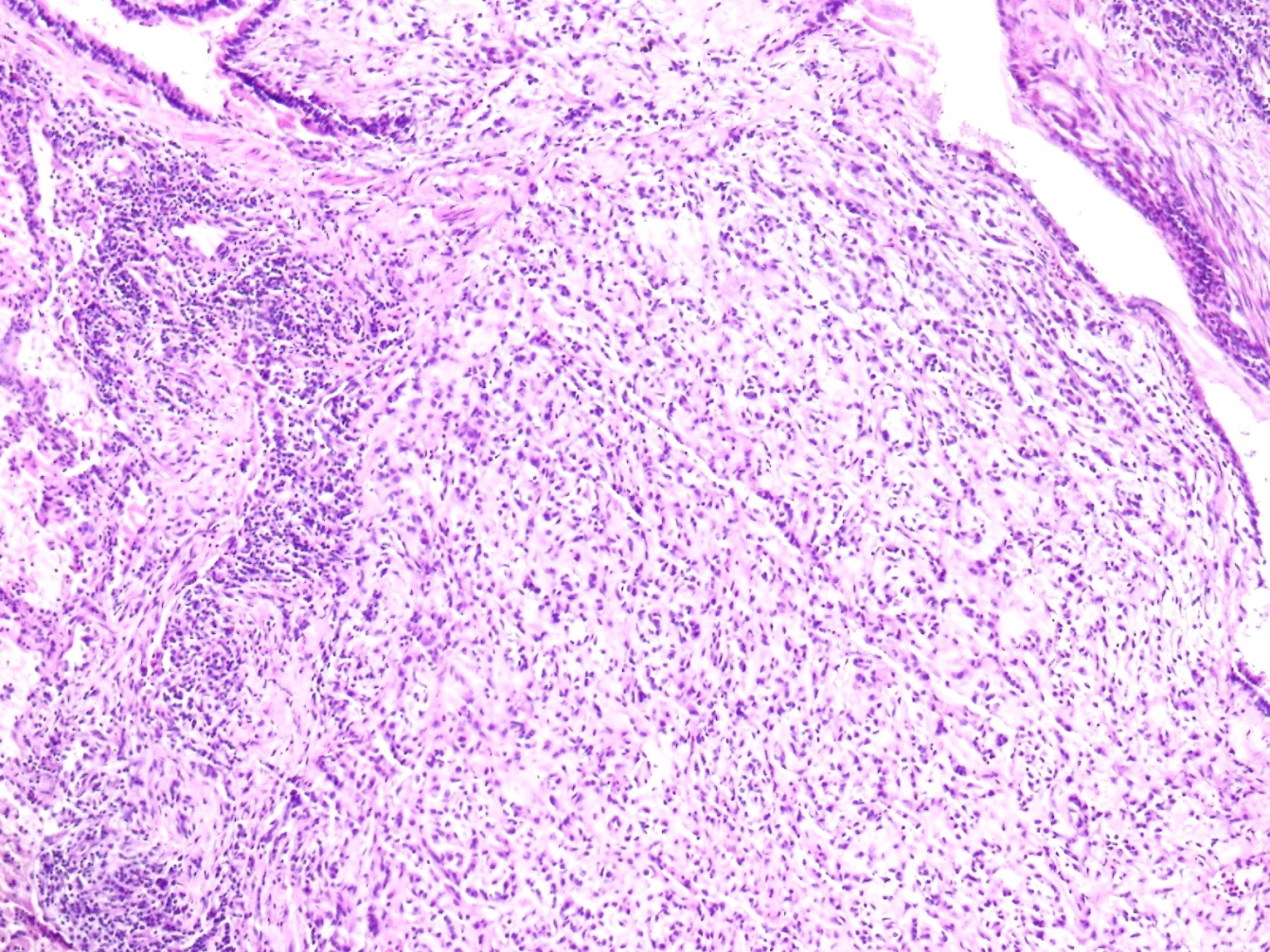
Figure 4: 10x10 HE Uniform, spindle-oval, tumor cell proliferation in the abutant myxoid stroma
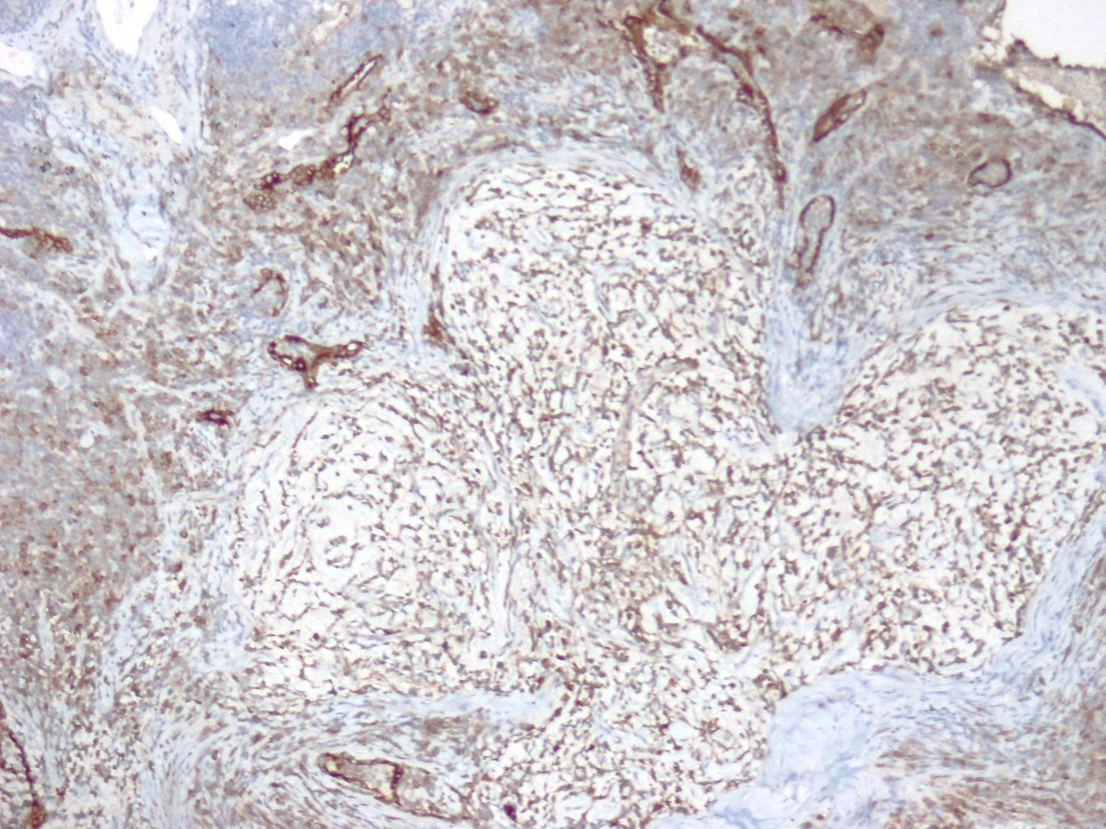
Figure 5: 10x10 EMA focal expression pattern
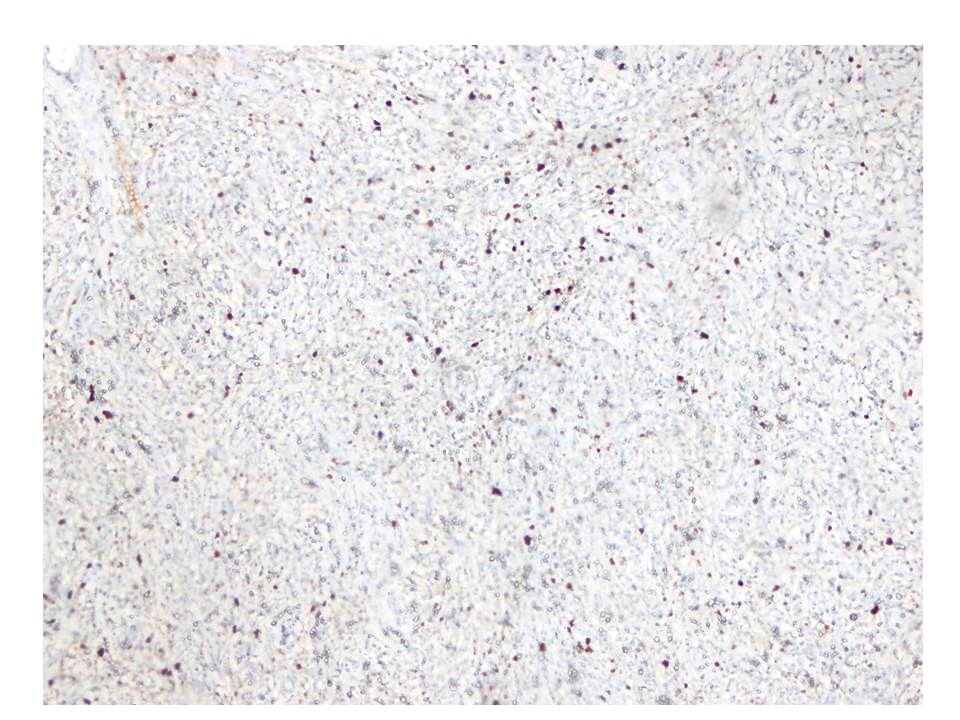
Figure 6: The Ki67 proliferation index was low
Figures at a glance