Breast Cancer Following Prophylactic Mastectomy in Mutation Carriers: Can a Retropectoral Reconstruction Make a Difference on Early Recognition? a Case Series
Received Date: December 09, 2022 Accepted Date: January 09, 2023 Published Date: January 11, 2023
doi: 10.17303/joo.2023.2.101
Citation: Cristina Garusi, Valeria Navach, Bernardo Bonanni, Eleonora De Antoni (2023) Breast Cancer Following Prophylactic Mastectomy in Mutation Carriers: Can a Retropectoral Reconstruction Make a Difference on Early Recognition? a Case Series. J Oncogenomics and Oncotarget 2: 1-8
Abstract
Background: BRCA1 and BRCA2 mutation carriers represent high-risk subjects who can benefit from prophylactic risk-reducing mastectomy. No mastectomy can remove all breast glands and consequently eliminate all risk, and the so-called “Angelina Jolie effect” requires plastic surgeons to perform the most cosmetic and immediate surgery. There are no guidelines indicating the preferred type of reconstruction following a prophylactic mastectomy; the only concern is for the type of mastectomy and the indication for an immediate reconstruction. The literature reports extremely rare cases of invasive breast cancer in previously risk-reducing mastectomies for mutation carriers.
Case description: We present two cases of breast cancer following prophylactic mastectomy and retropectoral implant reconstruction that were easily detected and treated.
Discussion: In the light of the current tendency towards simple clinical surveillance and prepectoral implant reconstructions, we discuss the oncological benefit of the retromuscular plane in high-risk patients.
Keywords: BRCA Mutation Carrier; Risk-Reducing Mastectomy; Prophylactic Mastectomy; Breast Reconstruction
Introduction
Although most breast and ovarian cancers are sporadic, the percentage of inherited breast cancer is 7-8%, while that for ovarian cancer ranges from 15-25%.
About a quarter of inherited breast cancers are due to BRCA1 and BRCA 2 mutation carriers.
In BRCA 1 mutation carriers, the risk of developing breast cancer during their lifetime may vary between 35% and 80%, while in BRCA 2 mutation carriers this may vary between 30% and 65% [1].
BRCA 1 mutation carriers have a higher risk for breast and ovarian cancer compared to the general population. BRCA 2 mutation carriers have a higher risk in developing other cancers such as melanoma, and those of the pancreas, gastrointestinal system, prostate and breast in the male population, and breast and ovaries in the female population [1].
Regarding breast cancer, in BRCA1 the risk of a triple-negative tumour is higher. Today the possibility of survival after breast cancer is 80% at 5 years if early diagnosis is performed. The incidence in Italy is about 55,000 new cases per year with a prevalence of 843,200 Italian women who are surviving despite breast cancer. Male breast cancer is about 8-10 times higher compared to non-mutated men [1].
Ovarian cancer is one of the “big killers”, with early diagnosis being especially difficult.
With the most recent treatments the possibility of survival at 5 years is now 40-50% after diagnosis. The expected incidence is 5,200 new cases per year in Italy, with a prevalence of 49,800 Italian women surviving after ovarian cancer. The ovarian cancer lifetime risk is about 25-40%for BRCA1 and BRCA2 patients, with a later onset for BRCA2 patients, so ovariectomy is indicated by 40 years of age for BRCA1 and between 40 and 45 years of age for BRCA2 [1]
To reduce the risk of breast cancer, bilateral prophylactic mastectomy represents the surgical option, generally preferred to enhanced radiological surveillance and hormone-therapy. Although studies show a 90% risk reduction after surgery in BRCA carriers, the risk can never totally be eliminated because efficacy is likely linked to the residual breast tissue [1].
Some terminal duct lobular units may remain with skin flap thickness >5 mm, in the axillary extension and within the pectoralis fascia, and ducts survive in the nipple [2].
Over the years, mastectomy techniques have evolved from radical to simple mastectomy and have become more conservative with the so-called subcutaneous mastectomies (skin-sparing mastectomy SSM, nipple-sparing mastectomy NSM, and skin reducing mastectomy SRM) [3], which are oncological safe and allow for improved aesthetic and functional outcomes. Every mastectomy removes the pectoralis fascia, and subcutaneous mastectomies in particular have anterior dissection just above the superficial fascia, which is sometimes difficult to identify. In a subcutaneous mastectomy, the breast envelope and the inframammary fold are spared, ideally enabling less loss of sensation and better-restored final morphology through Immediate Breast Reconstruction (IBR).
It is reported in the published medical literature that the distance from the superficial fascia to skin is <1.1 mm in 50% of patients [4] and that residual breast tissue can be found in 58.3% of prophylactic mastectomies and in 51.6% of therapeutic nipple-sparing mastectomies [5].
According to AHS, CCO, and the NCCN guidelines, nipple-sparing mastectomy is appropriate for prophylactic mastectomy, and a recent review of breast reconstruction guidelines indicates that immediate reconstruction is recommended for women undergoing prophylactic mastectomy [6].
Reconstructive choice is both a surgeon and a patient’s decision, based upon many factors: eventual donor sites for autologous flaps or fat graft, risk factors, breast cup and ptosis, and the patient’s expectation and willingness.
Implant reconstruction is the most performed breast reconstruction worldwide. Many options are available: a total retromuscular pocket; a “dual plane” pocket where the implant is placed both behind the pectoralis major muscle on the superior-internal side and behind the mastectomy skin flap with or without an ADM/mesh on the lower-external side; and a new prepectoral position right behind the mastectomy flap, covered by an ADM or a polyurethane shell.
The location of the pocket is determined by the vitality of the mastectomy flap: a prepectoral implant pushing over a poor skin flap risks complications (ischemia, dehiscence, infection, extrusion) so it is only connected to a thick, well-perfused mastectomy flap. Dual plane and retromuscular reconstruction, instead, reduce implant pressure over the mastectomy flap and can be approached with “skinny” mastectomy flaps. Total and partial retropectoral reconstruction may cause more postoperative pain, muscle spasm and hyper-animation, while prepectoral reconstruction may offer natural breast shape, lower capsular contracture and greater satisfaction with outcomes [7].
The prophylactic primum movens of mastectomy in healthy mutation carrier’s demands both the most complete breast tissue removal and the most cosmetic result; it is a surgical primary prevention where oncological and aesthetic criteria must be embraced, but this union is not simple to achieve. In particular, the thickness of the mastectomy flap should not be decided based upon the type of the aimed reconstruction, but the reconstruction should be decided according to the mastectomy flap. Few reports in the literature cite the event of breast cancer after prophylactic mastectomy for BRCA mutation carriers [8,9].
We reported two additional clinical cases of breast cancer following prophylactic mastectomy in BRCA mutation carriers who were treated at the IEO, European Institute of Oncology IRCSS, in Milan, Italy.
Case presentation
All procedures were performed in accordance with the ethical standards of the institutional and national research committees and with the Helsinki Declaration (as revised in 2013).
The first case involved a 34-year-old female BRCA1 mutation carrier, who had a clinically palpable 2cm subcutaneous cancer on a 3-year-old left prophylactic nipple-sparing mastectomy reconstructed with a total retropectoral implant (Figure 1a). When she was 31 years old in 2015, she underwent surgery, after having undergone a CT (EC x 4, taxol x 3), for triple-negative tumor of her right breast with nipple-sparing mastectomy, sentinel node dissection and immediate implant reconstruction. The final histology was: pT1c(m) (1.5 cm), pNSent Neg (0/3), G3, vascular invasion absents, receptors ER 0%, PgR 0%, Ki-67 60%, c-erbB2: absent.
Genetic consultation found her to be BRCA1 positive.
It was therefore suggested, in 2016, at the age of 32, that she undergo risk-reducing left side mastectomy and immediate implant reconstruction with the same total muscle-covered implant as the contralateral side.
In 2018 a nodule of 2cm was diagnosed in the healthy side with concomitant 6 mm nodule on the right side and capitation at PET scan in the right mastectomy skin and right lung (suspected metastasis). She was treated with chemotherapy with carboplatin and gemcitabine, then changed to cisplatin.
At the end of the treatment, in 2019 she underwent wide bilateral excision, and bilateral change of implants to expanders.
The histology was positive only on the left breast (previous healthy side). Staging of the left breast was ypT1a (3 mm); ypN0(sentinel); negative receptors.
She underwent removal of the single lung metastasis with a previous Technetium intralesional injection and subsequent change of expanders to implant (Figure. 1b).
In the second case, a 45-year-old female BRCA2 mutation carrier, was found at ultrasound (US) with a 9mm subcutaneous cancer marginally to the sternum in a 6-year-old prophylactic nipple -sparing right mastectomy with total sub-muscular reconstruction.
At 39 years old, she had been diagnosed with infiltrating breast cancer of her left breast. She underwent an immediate genetic test that revealed BRCA2 mutation.
She therefore underwent, in 2015, left nipple-sparing mastectomy, sentinel node biopsy and risk-reducing right side and bilateral total sub-muscular implant reconstructions.
The histology was positive in the left side: cribriform well-differentiated breast cancer, with large intraductal component pT1c (1.8 cm) pNSent Neg (0/3), G1, IVP absent, ER 95% PgR 95%, Ki-67 8%, c-erbB2: partial weak in 90%, Ca 15-3: 23 u/ml.
She was treated with tamoxifen 20 mg/die until 11/2020.
In 06/2018 she underwent laparoscopic ovariectomy with negative histology.
All remained stable until January 2021 when ultrasound revealed a dense lesion of 8 - 9 mm on the right breast, QII, marginally to the sternum.
After a straightforward fine needle aspiration (FNA) (Figure 2a), wide excision of the nodule, sentinel node biopsy and bilateral implant change were performed (Figure. 2b).
Dicussion
According to the published medical literature, breast reconstruction for breast cancer, either with implants or autologous tissue, does not modify the risk of breast cancer recurrence compared to conventional mastectomy without reconstruction [10-12], and the breast cancer outcome is the same with alloplastic and autologous reconstruction [13]. A recent systematic review on the pattern of local recurrence after therapeutic SSM and NSM mastectomy and reconstruction with implants and flaps reports a 4.7% local relapse rate: skin and/or subcutaneous tissue recurrence was 75.8%, chest wall was 14.2%, and NAC was in 10% of the cases [14].
The European Society for Medical Oncology (ESMO) guidelines recommend periodic clinical follow-up for operated early breast cancer; mammography is recommended only for breast conservative surgery and contralateral side, while US and breast MRI should be performed only when needed [1]. No specific indication is made for locally advanced breast cancer survivors,nor for surveillance of BRCA mutation carriers who have undergone risk-reducing mastectomy [1]. Clinical examination is the primary evaluation in every post-mastectomy breast, with radiologic examinations required only when there is clinical suspicion.
The appropriate surveillance imaging strategy is a debated topic [15], with evidence from small retrospective studies. In 2020, the American College of Radiology (ACR) published Appropriateness Criteria Imaging After Mastectomy and Breast Reconstruction by a panel of experts [16]. According to the ACR, in high-risk patients with prophylactic mastectomy and autologous or implant reconstructions and absence of clinical suspicion, there is insufficient evidence to support the use of mammography, digital breast tomosynthesis and US; although MRI may be useful in monitoring the residual breast tissue, there is insufficient evidence to support its use; it is suggested that the yield of screening is especially low in the setting of retropectoral implant placement, in which recurrences are most likely to be clinically palpable.
Breast reconstruction with autologous tissues is typically performed in a prepectoral location because there is no need to lift the pectoralis major muscle. In this setting, the clinical detection of cancer may suffer from frequent partial fat necrosis and oil cysts, and radiological follow-up is often performed to exclude benign lesions; adipose tissue of flaps allows for improved contrast with dense lesions such as cancer nodules on mammography.
In an implant-based reconstruction, mastectomy boundaries are different for each pocket plane; in a total retromuscular reconstruction, the breast bed is totally subcutaneous; in a partial retromuscular reconstruction, it is both subcutaneous and behind the lower-external part of the implant; in a prepectoral reconstruction, it is both subcutaneous and behind the whole implant. Cancer relapses both in the superficial plane and on the chest wall have been reported in the published medical literature, but no distinction is made regarding implant location [14].
No trial has ever investigated cancer occurrence in previous prophylactic mastectomies, and no report up to this day has investigated incidences of cancer occurrence comparing different reconstructions after risk-reducing mastectomy. A recent original article on prepectoral implant reconstruction for prophylactic mastectomy reported that no patients developed breast cancer during 18 months of follow-up [17]. However, prepectoral reconstruction is a relatively new technique and it could be that this type of evidence has yet to be demonstrated. No literature exists regarding specific different radiological follow-up cautions associated with prepectoral implant reconstruction, while the European Society for Radiation and Oncology - Advisory Committee on Radiation Oncology Practice already has proper indications on radiotherapy [18].
From an anatomical point of view, circumscribing the mastectomy boundaries and its bed to a subcutaneous plane as in retropectoral implant reconstruction may help the clinician to conduct a local examination and, if need be, to easily perform US and FNA. In a prepectoral reconstruction, a tumor could occur behind the implant and potentially be identified later, and a posterior biopsy would probably require implant removal and major surgery.
The prepectoral solution is undoubtedly a great innovation and yields optimal results; particularly in these “Angelina Jolie effect” times, a prepectoral implant reconstruction is a solution for the aesthetically demanding and very young healthy female population. A specific proper radiological follow-up should be encouraged anyway, as mutation carriers still have breast tissue and still harbour the potential for an aggressive cancer; additional studies should investigate the oncologic outcome between different reconstructions over different pocket planes and the appropriate surveillance for every case should be recommended
Women need to be aware that prophylactic mastectomy can reduce the risk of breast cancer without completely eliminating it. Close collaborations between breast surgeons, breast radiologists and plastic surgeons can indicate the correct surgical treatment for mastectomy, reconstruction, and radiological surveillance. A careful follow up based on clinical examination remains necessary, as delayed cancer can occur. Risk-reducing mastectomy in high-risk patients could be associated with total submuscular reconstruction for simpler follow-up, or with prepectoral reconstruction for women with aesthetic expectations who understand the need for thorough whole-breast-bed surveillance.
Footnote
The authors declare that they have no conflict of interest nor funding to disclose.
The authors are accountable for all aspects of the work in ensuring that questions related to the accuracy or integrity of any part of the work are appropriately investigated and resolved.
This article does not contain any studies with human participants or animals performed by any of the authors. All procedures performed in this study were in accordance with the ethical standards of the institutional and/or national research committee(s) and with the Helsinki Declaration (as revised in 2013). Written informed consent was obtained from the patient for publication of this case report and accompanying images. A copy of the written consent is available for review by the editorial office of this journal.
Conflict of interest
none
Funding
none
- Garusi C, Bonanni B, Peccatori F, Morelli F (2021) Scacco ai geni. Techiche nuove.
- Torresan RZ, Dos Santos CC, Okamura H, Alvarenga M (2005) Evaluation of residual glandular tissue after skin-sparing mastectomies. Ann Surg Oncol 12: 1037-44.
- Matthew D Freeman, Jared M Gopman, C Andrew Salzberg (2018) The evolution of mastectomy surgical technique: from mutilation to medicine. Gland Surg 7: 308-15.
- Beer GM, Varga Z, Budi S, Seifert B, Meyer VE (2002) Incidence of the superficial fascia and its relevance in skin-sparing mastectomy. Cancer 94: 1619-25.
- Gregolin Giannotti D, Abdallah Hanna S, Cerri GG, Barbosa Bevilacqua JL (2018) Analysis of Skin Flap Thickness and Residual Breast Tissue After Mastectomy. J Radiat Oncol Biol Phys 102: 82-91.
- Popowich B, Kostaras X, Temple-Oberle C (2020) Breast reconstruction after therapeutic or prophylactic mastectomy for breast cancer: A comparison of guideline recommendations. Eur J Surg Oncol 46: 1046-51.
- Cuomo R (2020) Submuscular and Pre-pectoral ADM Assisted Immediate Breast Reconstruction: A Literature Review. Medicina (Kaunas) 56: 256.
- Maarse W, Jonasse Y, Ausems MG, Schipper ME, van Hillegersberg R (2009) First case of invasive breast cancer following prophylactic bilateral skin sparing mastectomy in a BRCA1 mutation carrier. Eur J Surg Oncol 35: 1016-8.
- Kasprzak L, Mesurolle B, Tremblay F, Galvez M, Halwani F et al. (2005) Invasive breast cancer following bilateral subcutaneous mastectomy in a BRCA2 mutation carrier: a case report and review of the literature. World J Surg Oncol 3: 52.
- Eriksen C, Frisell J, Wickman M, Lidbrink E, Krawiec K et al. (2011) Immediate reconstruc-tion with implants in women with invasive breast cancer does not affect oncological safety in a matched cohort study. Breast Cancer Res Treat 127: 439-46.
- McCarthy CM et al. (2008) Breast cancer recurrence following prosthetic, post- mastectomy recon-struction: incidence, detection, and treatment. Plast Reconstr Surg 121: 381-8.
- Geers J et al. (2018) Oncological safety of autologous breast reconstruction after mastectomy for invasive breast cancer. BMC Cancer 18: 994
- Zhen‐Yu W et al. (2022) Breast cancer outcomes following immediate breast reconstruction with implants versus autologous flaps: a propensity score‐matched study. Breast Cancer Res Treat191: 365-73.
- Joo JH (2021) Pattern of local recurrence after mastectomy and reconstruction in breast cancer pa-tients: a systematic review. Gland Surg 10: 2037-46.
- Trop I (2018) Is There a Role for Imaging Surveillance after Mastectomy and Autologous Breast Reconstruction? Radiology 289: 49-50.
- Heller SL et al. (2020) ACR Appropriateness Criteria Imaging After Mastectomy and Breast Re-construction. J Am Coll Radiol 17: S403-S14.
- Maruccia M et al. (2021) Prepectoral breast reconstruction: an ideal approach to bilateral risk-reducing mastectomy. Gland Surg 10: 2997-3006.
- Kaidar-Person O et al. (2019) ESTRO ACROP consensus guideline for target volume delineation in the setting of postmastectomy radiation therapy after implant-based immediate reconstruction for early stage breast cancer. Radiother Oncol 137: 159-66.
FIGURE 1
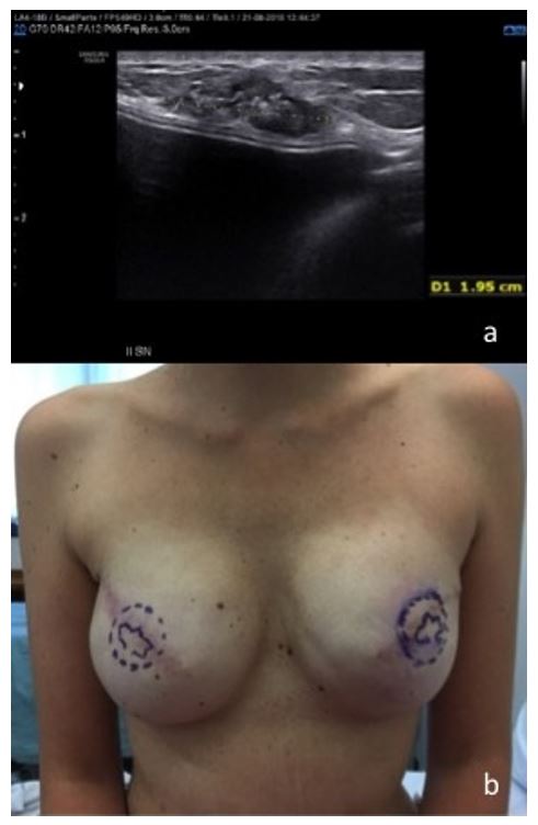
Figure 1: (a) Case 1. US view of subcutaneous cancer on a 3-year-old left prophylactic nipple-sparing mastectomy reconstructed with total retropectoral implant in a BRCA1 mutation carrier. (b) After bilateral wide excisions (including the nipple-areola complexes) of the left cancer and right suspect nodule, the patient underwent bilateral expander reconstruction and subsequent exchange with implants; in this photograph, the patient is awaiting nipple-areola reconstruction.
FIGURE 2
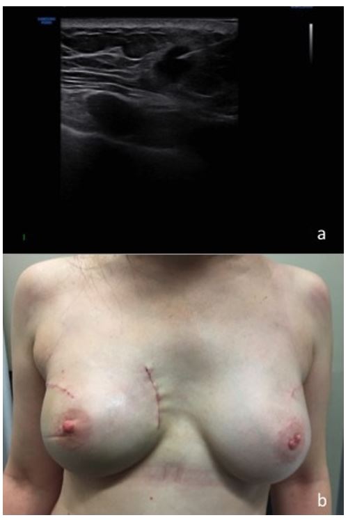
Figure 2: (a) Case 2. US view of FNA of subcutaneous cancer in a 6-year-old prophylactic nipple-sparing right mastectomy with total submuscular reconstruction in BRCA2 mutation carrier. (b) Postoperative view after wide excision of the right cancer, sentinel node biopsy and bilateral submuscular implant change.
Figures at a glance