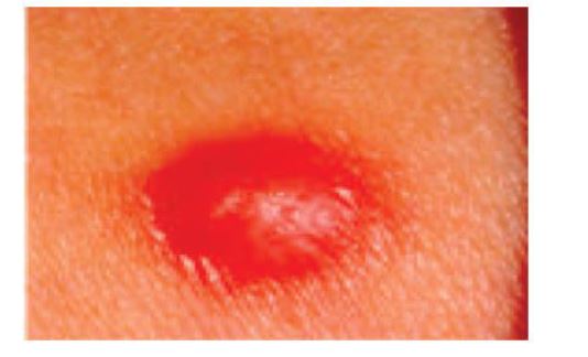
Figure 1: Clinical image of the firm brown-pink smooth papule on the right upper arm (florid leiomyoma)
Figure 1: Clinical image of the firm brown-pink smooth papule on the right upper arm (florid leiomyoma)
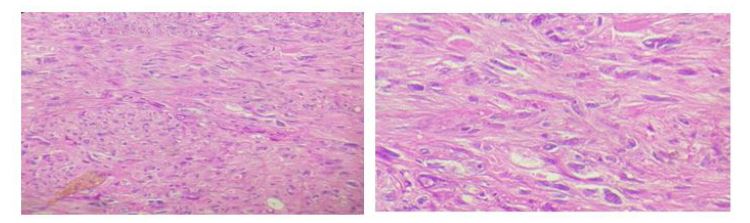
Figure 2: Histologically, the lesion showing long bundles of large spindle cells with a nuclear pleomorphism and without mitotic figures. (Haematoxylin and eosin stain (1Ox) &(40x))
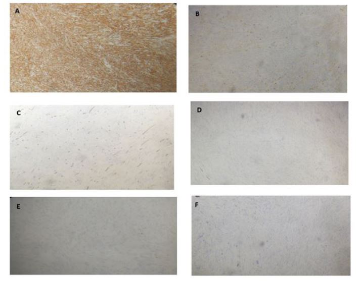
Figure 3: Immunohistochemical studies showing positive staining of H-caldesmon (A) and desmin(B) with negative staining of CD34 (C), pancytokeratin (D) , S100 (E) and a weak index of cell proliferation Ki 67 estimated at 2 percent (F)
Figures at a glance