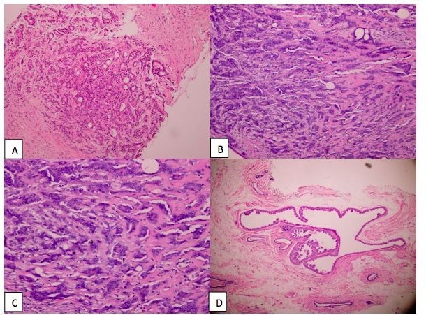
Figure 1a: Tubular proliferation showing proliferative glandular structures bordered by a double layer of epithelial and myoepithelial cells (Haematoxylin and eosin x100)
Figure 1b: Proliferative glandular structures bordered by epithelial and myoepithelial cells within a central area of dense scar-like fibrosis. (Haematoxylin and eosin x100)
Figure 1c: The stroma was fibrous and abundant compressing the epithelial structures and reducing their lumen giving an appearance of pseudo-infiltrative streaks. (Haematoxylin and eosin x200)
Figure 1d: Adjacent breast parenchyma exhibited apocrine metaplasia (Haematoxylin and eosin x100)
Tables at a glance
Figures at a glance