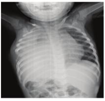
Figure 1: opacity of the left hemithorax compressing heart and the mediastinum structures to the right
Figure 1: opacity of the left hemithorax compressing heart and the mediastinum structures to the right
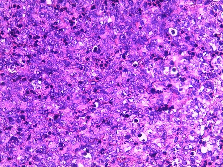
Figure 2: Eosine staining showing large cell ranges with often poorly reduced cytoplasm and oval nuclei with vesicular chromatin, and cell ranges with nuclei often eccentrically placed with abundant eosinophilic cytoplasmic inclusion
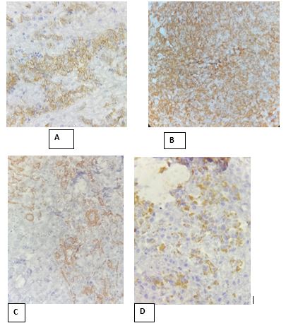
Figure 3: A: Vimentin, B: CD56, C: SMA, D: CKAE1/AE3
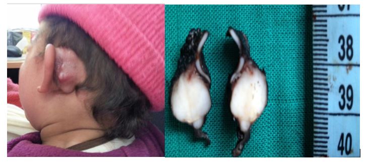
Figure 4: left retro auricular mass
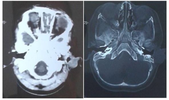
Figure 5: Retro auricular soft tissue process without endocranial extension
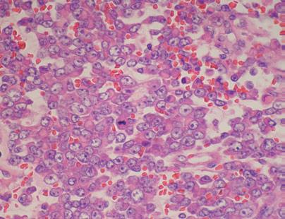
Figure 6: Hematoxylin eosin staining showing atypical rhabdoide cells with vesicular nucleolus nuclei and eosinophilic intra cytoplasmic inclusion
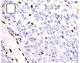
Figure 7: Negative staining with INI1
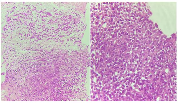
Figure 8: Proliferation of round cells with an abundant eosinophilic cytoplasm and a rounded or oval hyperchromatic and eccentrically placed nucleus arranged in plaques on a myxoid background
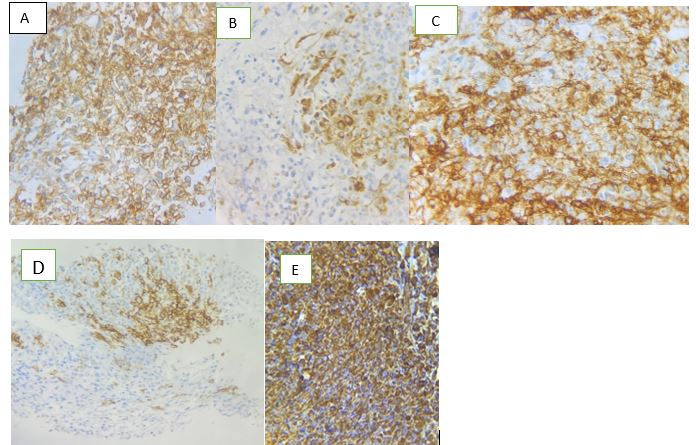
Figure 9: A= CD99+; B=CKAE1/AE3+; C=EMA+; D=CD56+; E= Vimentin+
Figures at a glance