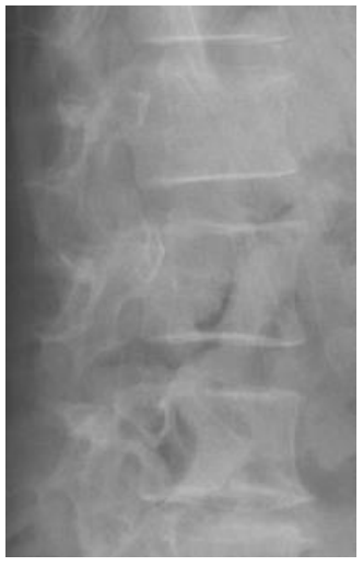
Figure 1: X-ray of the lumbar spine in lateral projection without valuable findings
Total |
Mean ± SD or n (%) |
|
Gender |
Male |
31 (68.22) |
FeMale |
14 (37.78) |
|
Smoker |
Never |
16 (35.55) |
Former smoker / smoker |
29 (64.45) |
|
Pattern |
Axial |
8 (17.77) |
Mixed |
37 (82.23) |
|
BMI |
Normal |
19 (42.22) |
Overweight / obese |
26 (57.78) |
|
MASES baseline |
|
1.3 ± 1.73 |
Dactilytis baseline (yes / no) (n) |
|
14 / 31 |
PASI baseline |
|
2.58 ± 1.96 |
NSAID |
Continuous |
9 (20) |
On demand |
36 (80) |
|
bDMARDs |
Yes |
18 (40) |
No |
27 (60) |
|
HLA-B27 |
Positive |
10 (22.22) |
|
Negative |
35 (77.78) |
CRP mean (unit) |
|
0.63 ± 0.45 |
ASDAS-CRP mean (unit) |
|
1,72 ± 0,61 |
BASFI baseline/ BASFI final |
|
3.42 ± 2.30 / 2.97 ± 2.12 |
BASMI 3 baseline/ BASMI 3 |
|
2.48 ± 2.18 / 2.52 ± 2.12 |
PASRI baseline/ PASRI final |
|
10.46 ± 8.40 / 12.28 ± 10.35 |
Fracture * (yes / no) (n) |
|
13 / 32 |
MASEI (baseline) |
|
16.35 ± 10.39 |
Table 1: Baseline characteristics of the patients
Abbreviations: ASDAS-CRP: Ankylosing Spondylitis Disease Activity Score-PCR; bDMARDS; biological disease-modifying antirheumatic drug; BASFI: Bath Ankylosing Spondylitis Functional Index; BASMI: Bath Ankylosing Spondylitis Metrology Index; BMI: body mass index; CRP: C Reactive Protein; MASES: Maastricht Ankylosing Spondylitis Enthesitis Score; NSAID: non-steroid anti-inflammatory drug; PASI: Psoriasis Area Severity Index; MASEI: Madrid Sonographic Enthesitis Index; PASRI: Psoriatic Arthritis Spondylitis Radiology Index.
Relationship between variables and radiographic progression: Male gender, smoking and patients with vertebral fracture were factors associated with greater radiographic progression. The relationship between the qualitative variables with radiographic progression is shown in table 2.
|
Mean (SD) |
p |
Gender (male / female) |
3.00 (2.47) / 1.42 (1.22) |
* |
Smoker (yes / no) |
3.68 (2.65) / 1.86 (1.76) |
* |
Axial vs mixed |
1.87 (1.45) / 2.64 (2.40) |
** |
Normal vs overweight/obese |
9.73 (10.43) / 14.15 (10.08) |
** |
Dactilytis (yes / no) |
2.64 (2.53) / 2.45 (2.18) |
** |
NSAID dosing (+) |
1.55 (1.33) / 2.75 (2.40) |
** |
bDMARDs (yes / no) |
1.83 (1.61) / 2.96 (2.54) |
** |
HLA-B27 (positive / negative) |
2.12 (1.64) / 2.57 (2.31) |
** |
Vertebral fracture (yes / no) |
4.00 (2.57) / 1.83 (1.79) |
* |
Abbreviations: NSAID: non-steroid anti-inflammatory drug; (+) continuous / on demand; * p < 0,05, **N.S.
Table 2: Relationship between baseline characteristics and radiographic progression
Variables |
R |
P value |
Age |
0.18 |
0.2 |
Time of evolution |
0.19 |
0.2 |
BMI |
0.29 |
0.05 |
Modified MASES |
0.04 |
0.7 |
ASDAS-CRP |
0.27 |
0.06 |
Baseline BASFI |
-0.01 |
0.9 |
Differential BASFI |
0.22 |
0.1 |
Baseline BASMI |
0.09 |
0.5 |
Differential BASMI |
-0.11 |
0.9 |
PASI |
-0.09 |
0.5 |
CRP |
0.2 |
0.1 |
Baseline PASRI |
0.49 |
0.001 ** |
Total MASEI |
0.35 |
0.01 * |
Calcification |
0.36 |
0.01 * |
Structure |
0.32 |
0.02 * |
Thickening |
0.11 |
0.43 |
Bursitis |
-0.27 |
0.07 |
Erosion |
0.14 |
0.34 |
Power-doppler |
0.02 |
0.89 |
Abbreviations: ASDAS-CRP: Ankylosing Spondylitis Disease Activity Score-PCR; BASFI: Bath Ankylosing Spondylitis Functional Index; BASMI: Bath Ankylosing Spondylitis Metrology Index; BMI: body mass index; CRP: C Reactive Protein; MASES: Maastricht Ankylosing Spondylitis Enthesitis Score; PASI: Psoriasis Area Severity Index; MASEI: Madrid Sonographic Enthesitis Index; PASRI: Psoriatic Arthritis Spondylitis Radiology Index.
Table 3: Correlation coefficients between clinical, radiographic and ultrasound variables and radiographic progression
Variable |
β |
Confidence interval |
P value |
R2 |
Male gender |
1.4 |
0.07, 2.73 |
0.04 |
0.23 |
Smoking |
1.63 |
0.40, 2.97 |
0,01 |
|
Vertebral fracture |
1.84 |
0.48, 3.05 |
0.004 |
0.37 |
Baseline PASRI |
0.42 |
0.04, 0.18 |
0.001 |
|
Calcification |
0.36 |
0.03, 0.28 |
0.01 |
0.12 |
Abbreviations: PASRI: Psoriatic Arthritis Spondylitis Radiology Index.
Relationship between variables in patients who did not have radiographic progression: Twelve patients had no radiographic progression (26.7%). These patients had a lower PASRI at baseline (5.25 + 2.63 vs 12.36 + 8.99; p = 0.004). No differences were found in the rest of the variables analysed (table 5).
Table 4: Multiple linear regressions by steps between the radiographic progression and clinical variables, radiographic damage and initial ul- trasound damage variables
|
No radiographic progression |
Radiographic progression |
p value |
n (%) |
12 (26.7) |
|
|
Gender male / female (%) |
22.6 |
35.7 |
0.3 |
Age |
51.67 ± 14.09 |
54.18 ± 12.64 |
0.5 |
Smoker / non-smoker (%) |
12.5 |
34.5 |
0.15 |
Pattern of involvement (%) axial / mixed |
25 |
27 |
0.91 |
BMI |
26.12 ± 4.46 |
28.09 ± 4.66 |
0.14 |
MASES |
2.00 ± 1.95 |
1.15 ± 1.67 |
0.13 |
NSAID dosing (%) continuous / on demand |
22.2 |
27.8 |
0.76 |
bDMARDs (%) yes / no |
33.3 |
22.2 |
0.45 |
HLA B27 (%) positive /negative |
30 |
25.7 |
0.96 |
CRP MEDIA |
0.49 ± 0.27 |
0.68 ± 0.50 |
0.35 |
Dactylitis (%) yes / no |
28.6 |
25.8 |
0.84 |
Vertebral fracture (%) Yes (or) No |
14.3 |
32.3 |
0.2 |
Total MASEI |
14.92 ± 10.44 |
16.82 ± 10.05 |
0.43 |
Calcification |
5.00 ± 4.65 |
7.09 ± 5.37 |
0.22 |
Structure |
2.08 ± 2.96 |
2.94 ± 3.08 |
0.11 |
Thickening |
0.92 ± 0.90 |
1.15 ± 1.48 |
0.92 |
Bursitis |
1.17 ± 0,71 |
0.73 ± 0.83 |
0.15 |
Erosion |
3.75 ± 5.13 |
3.15 ± 3.43 |
0.91 |
Power-doppler |
2.00 ± 1.95 |
1.55 ± 1.85 |
0.55 |
ASDAS-CPR (mean) |
1.78 ± 0.59 |
1.53 ± 0.65 |
0.08 |
BASFI baseline |
3.10 ± 2.15 |
3.54 ± 2.37 |
0.6 |
BASFI differential |
0.51 ± 3.01 |
-0.28 ± 2.36 |
0.79 |
Baseline BASMI |
1.83 ± 0.93 |
2.72 ± 2.33 |
0.4 |
BASMI 4 differential |
0.06 ± 0.34 |
0.00 ± 0.42 |
0.79 |
PASI |
3.44 ± 2.03 |
2.26 ± 1.83 |
0.05 |
Baseline PASRI |
5.25 ± 2,63 |
12.36 ± 8.99 |
0.004 |
Abbreviations: ASDAS-CRP: Ankylosing Spondylitis Disease Activity Score-PCR; bDMARDS; biological disease-modifying antirheumatic drug; BASFI: Bath Ankylosing Spondylitis Functional Index; BASMI: Bath Ankylosing Spondylitis Metrology Index; BMI: body mass index; CRP: C Reactive Protein; MASES: Maastricht Ankylosing Spondylitis Enthesitis Score; NSAID: non-steroid anti-inflammatory drug; PASI: Psoriasis Area Severity Index; MASEI: Madrid Sonographic Enthesitis Index; PASRI: Psoriatic Arthritis Spondylitis Radiology Index.
Table 5: Relationship between variables in patients who did not have radiographic progression
Figure 1: X-ray of the lumbar spine in lateral projection without valuable findings
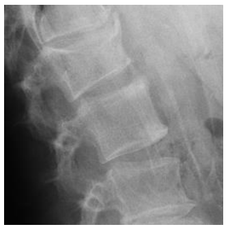
Figure 2: X-ray of the lumbar spine in lateral projection in the same patient with loss of height and vertebral wedging and the formation of a syndesmophyte
Tables at a glance
Figures at a glance