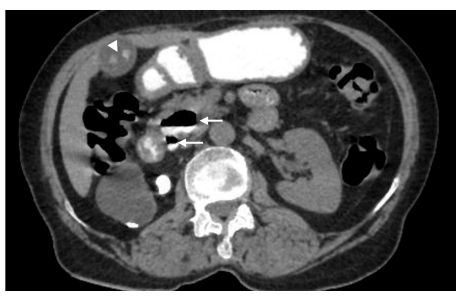
Figure 1: NCCT upper abdomen with oral contrast. It shows two contrast- lled diverticula (white arrows) with an air- uid (contrast) level.
Also, note hyperdense calculi (white arrowhead) in the lumen of the gall bladder
FIGURE 2
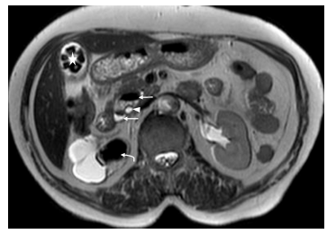
Figure 2: MRCP. T2 weighted axial image shows two periampullary diverticula (white arrows) around the common bile duct (white arrowhead) causing narrowing of the distal common bile duct with a luminal diameter of 2.8 mm at this level. Also, note the cholelithiasis (white
star). Incidental nding of a large obstructive calculus (white curved arrow) in the right renal pelvis, which is causing gross hydronephrosis
FIGURE 3
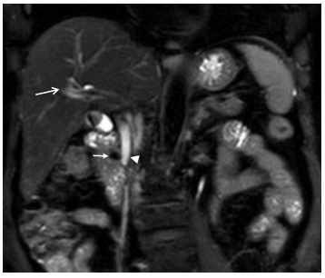
Figure 3: MRCP. T2-weighted SPAIR coronal image shows the smaller periampullary diverticulum (white arrow) with signi cant narrowing
of the distal common bile duct (arrowhead) with a luminal diameter of 2.8 mm at this level. Also, note the mild to moderate degree of intrahepatic biliary radical dilatation (long white arrow)
Figures at a glance