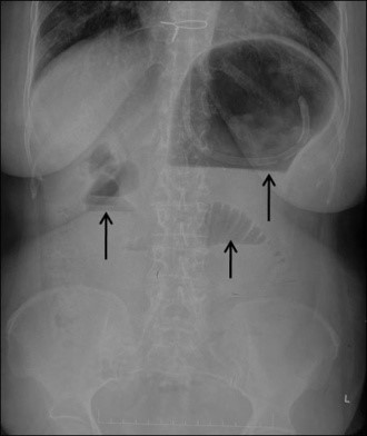
Figure 1: Plain radiograph of the abdomen in erect posture showing dilated stomach, duodenum, and the proximal jejunal loops with presence of air-fluid levels (black arrows).
Figure 1: Plain radiograph of the abdomen in erect posture showing dilated stomach, duodenum, and the proximal jejunal loops with presence of air-fluid levels (black arrows).
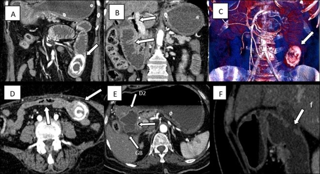
Figure 2(A-F):Contrast-enhanced CT images of the abdomen showing an ectopic gallstone impacted in proximal jejunum (A) with proximal dilated bowel and pneumobilia (B). Volume rendered CT image showing ectopic gall stone with dilated proximal small bowel(C). Bowel loops distal to the impacted gall stone are collpased (D) with delineation of the cholecysto-duodenal fistula (denoted by f) in the axial (E) and the sagittal (F) sections.
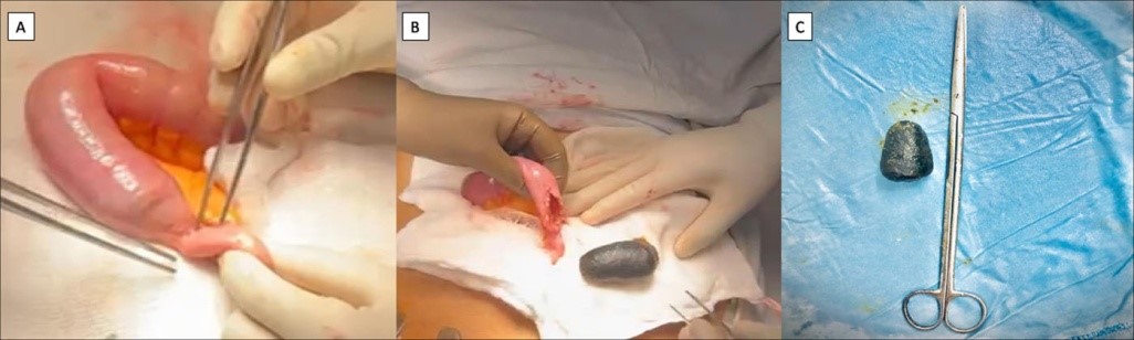
Figure 3(A-C): Intraoperative pictures showing impacted gall stone causing obstruction in proximal jejunum (A), longitudinal enterotomy with delivered stone (B) and retrieved black pigmented gall stone (C).
Figures at a glance