
Efficacy and Safety of Biomatrix Beat Sensor Versus ECG Gating in Cardiac MRI: A Systematic Review of Clinical and Technical Evidence
Received Date: July 13, 2026 Accepted Date: August 14, 2026 Published Date: August 18, 2026
doi:10.17303/jrnm.2026.3.103
Citation: Ajay Chand P, Deepthi Devadas, Pratheeksha Kamath, Bibin Joseph (2026) Efficacy and Safety of Biomatrix Beat Sensor Versus ECG Gating in Cardiac MRI: A Systematic Review of Clinical and Technical Evidence. J Radiol Nucl Med 3: 1-14
Abstract
Cardiac magnetic resonance imaging is often affected by motion artifacts caused by cardiac and respiratory cycles, which can reduce image quality and workflow efficiency. Conventional electrocardiogram gating is widely used for motion synchronization but may be limited by setup complexity, electromagnetic interference, and reduced reliability in patients with arrhythmias. This systematic review evaluated the efficacy, safety, and clinical utility of the BioMatrix Beat Sensor/Pilot Tone, a contactless technology integrated into MRI body coils for simultaneous cardiac and respiratory motion detection. A systematic search of PubMed, Scopus, Google Scholar, and Siemens Healthineers resources was conducted up to October 2025 following a PROSPERO registered protocol - (CRD420251121514). Due to limited evidence and study heterogeneity, a narrative synthesis was performed. Five primarily non-randomized studies and six studies from company resources were included. Pilot Tone triggering demonstrated quantitative CMR measurements comparable to ECG gating, including ventricular volumes, ejection fraction, strain, and T1/T2 mapping, with intraclass correlation coefficients ranging from 0.73 to 0.99. Image quality was similar between methods, while Pilot Tone showed advantages in patients with arrhythmias by reducing false triggers and motion artifacts. Respiratory signal correlation was high (r > 0.95). Workflow benefits included reduced preparation time of approximately 2–5 minutes and improved patient comfort due to the contactless design. No adverse events were reported. BioMatrix Beat Sensor/Pilot Tone appears to be a reliable alternative or complementary method to ECG gating in CMR, although larger multicentre studies are needed to confirm its broader clinical utility.
Keywords: Cardiac Magnetic Resonance Imaging (CMR); BioMatrix Beat Sensor; Pilot Tone; Electrocardiogram Gating (ECG); Motion Compensation; Arrhythmia; Image Quality; Respiratory Motion Tracking.
List of Abbreviations
MRI – Magnetic Resonance Imaging; CMR – Cardiac Magnetic Resonance; ECG – Electrocardiogram; PT – Pilot Tone; BM – BioMatrix; MRCP – Magnetic Resonance Cholangiopancreatography; MRA – Magnetic Resonance Angiography; EDV – End-Diastolic Volume; ESV – End-Systolic Volume; SV – Stroke Volume; EF – Ejection Fraction; LV – Left Ventricle / Left Ventricular; RV – Right Ventricle / Right Ventricular; SNR – Signal-to-Noise Ratio; CNR – Contrast-to-Noise Ratio; ICC – Intraclass Correlation Coefficient; CI – Confidence Interval; SD – Standard Deviation; CV – Coefficient of Variation; SG – Self-Gating; LGE – Late Gadolinium Enhancement; ECV – Extracellular Volume; BMI – Body Mass Index; SAR – Specific Absorption Rate; RF – Radiofrequency; MHD – Magnetohydrodynamic Effect; RCT – Randomized Controlled Trial; FDA – Food and Drug Administration; CE – Conformité Européenne (European Certification).
Introduction
Motion artifacts due to cardiac and respiratory cycles present significant challenges in magnetic resonance imaging (MRI), particularly in cardiovascular and abdominal imaging, where they can degrade image quality and extend examination times [1,2].Traditional synchronization methods, such as electrocardiogram (ECG) triggering and respiratory bellows or navigators, often encounter limitations including setup complexity, susceptibility to artifacts from magnetic fields, and variability in patient compliance [3,4]. Pilot Tone (PT) navigation, including its clinical application as the BioMatrix Beat Sensor, offers a contactless approach by transmitting a continuous radiofrequency signal modulated by physiological motion, enabling real-time extraction of respiratory curves and cardiac triggers independently of the MRI acquisition sequence [5,6].
The BioMatrix Beat Sensor, an innovative technology integrated into standard MRI body coils, eliminates the need for ECG leads and respiratory belts, simplifying patient preparation and reducing examination times [7,8]. This sensor leverages electromagnetic effects to detect cardiac and diaphragmatic motion, providing reliable triggering even in challenging cases such as arrhythmias, as demonstrated in clinical settings with improved image quality and workflow efficiency [9,10]. Its development, rooted in the Pilot Tone concept, has evolved from initial volunteer tests to a practical tool, enhancing patient comfort and diagnostic accuracy across various MRI scanners [7,10].
Recent studies have explored the efficacy of Pilot Tone and BioMatrix technologies across diverse clinical settings. In free breathing three-dimensional magnetic resonance cholangiopancreatography (MRCP), BioMatrix triggered imaging (BM MRCP) demonstrated reduced examination times and superior image quality compared to respiratory-gated and navigator-triggered techniques in patients with pancreatic and biliary diseases [1]. In cardiovascular MRI, Pilot Tone extracted signals showed high correlation with image-derived respiratory motion and reliable cardiac triggers, with minimal jitter relative to ECG, even across varying patient physiologies [2]. Initial clinical experiences at a regional cardiac centre highlighted the BioMatrix Beat Sensor’s ability to streamline workflows, reduce preparation time, and improve imaging quality in cardiomyopathy and aortopathy cases, particularly in patients with arrhythmias [3]. Furthermore, Pilot Tone integration into free running 5D flow MRI frameworks yielded respiratory and cardiac motion data comparable to self-gating methods, with the advantage of being acquisition independent(4). Prospective feasibility studies in patients with cardiovascular diseases confirmed that Pilot Tone triggered CMR matches ECG triggered imaging in quality and quantitative metrics, offering advantages in minimizing motion artifacts in cases of ECG unreliability [5]. Additionally, Pilot Tone triggered MRI showed good agreement with ECG triggered sequences for cardiac functional and structural indices, suggesting its potential as a backup gating method [6].
This systematic review synthesizes findings from these studies to evaluate the clinical performance, efficiency, and reliability of Pilot Tone and BioMatrix technologies in motion-resolved MRI, with a focus on their impact on patient outcomes and workflow optimization in cardiac and respiratory imaging.
Rationale for the Review
1.1.1 Methodological Rigour
Conventional ECG gating is frequently compromised by patient-specific factors (body size, chest hair, cardiac position, motion, arrhythmia) and electromagnetic interference from radiofrequency pulses and gradient switching, resulting in unreliable R wave detection and inconsistent cardiac synchronization. These limitations lead to motion artifacts, prolonged acquisition times, and increased rescans, reducing the reproducibility and diagnostic validity of cardiac MRI examinations. A systematic evaluation of contactless alternatives is essential to determine whether BioMatrix Beat Sensor/Pilot Tone consistently mitigates these well documented sources of error across varied clinical and technical conditions.
1.1.2 Clinical Credibility
In routine cardiology and radiology practice, ECG triggering remains particularly challenging in patients with pronounced arrhythmias like atrial fibrillation, frequent ectopic beats, implanted devices, or irregular breathing patterns, where electrode placement and signal stability directly impair image quality and quantitative accuracy. Clinical teams report that BioMatrix Beat Sensor eliminates ECG leads entirely, simplifies patient preparation, and delivers reliable triggering even in these difficult cohorts, resulting in fewer examination failures and more consistent diagnostic datasets suitable for cardiomyopathy, aortopathy, and structural heart disease assessment.
1.1.3 Evidence Strength
Individual reports demonstrate high agreement between Pilot Tone/BioMatrix signals and ECG/self-gating (correlation coefficients routinely >0.99), minimal trigger jitter, equivalent ventricular volumes, ejection fraction, strain, T1/T2 mapping values, and superior image quality in arrhythmia cases. However, this evidence is dispersed across single centre feasibility studies, clinical experience reports, and technical descriptions without formal aggregation. A systematic review is required to synthesize these findings, assess risk of bias (especially confounding and selection bias in non-randomized comparisons), and establish graded certainty of evidence to support clinical decision-making.
1.1.4 Innovation Viability
BioMatrix Beat Sensor is fully integrated into standard BioMatrix body coils, requires no additional hardware, and functions independently of acquisition sequence, making it immediately deployable on existing Siemens 1.5T and 3T platforms. By removing electrodes and respiratory belts, it substantially reduces preparation time, eliminates skin preparation and shaving, and improves patient tolerance advantages that translate directly into higher throughput and operational efficiency in high-volume cardiac MRI centres.
1.1.5 Audience Relevance
Radiologists, cardiologists, MRI technologists, and healthcare administrators routinely face workflow bottlenecks caused by ECG setup delays, signal instability, patient discomfort, and examination failures. These issues increase waiting times, rescheduling rates, and overall costs while compromising patient experience. This review directly addresses this audience by consolidating real world evidence on a commercially available, contactless solution that resolves longstanding practical and safety limitations in everyday cardiac MRI practice.
1.2 Deviations from PROSPERO Protocol
This review was prospectively registered in PROSPERO (CRD420251121514). During the conduct of the review, minor deviations from the registered protocol were required due to limitations in the available evidence.
Although the protocol planned to include randomized controlled trials, no randomized studies comparing BioMatrix Beat Sensor or Pilot Tone with ECG gating in cardiac MRI were identified. The available literature consisted of non-randomized comparative studies, feasibility studies, and clinical experience reports. Therefore, the inclusion criteria were broadened to incorporate these study designs in order to synthesize the best available clinical evidence.
Due to the small number of studies and substantial heterogeneity in study design, patient populations, imaging protocols, and outcome measures, a meta-analysis was not feasible. Accordingly, a structured narrative synthesis was performed, with descriptive reporting of outcomes without statistical pooling.
These deviations were driven by evidence availability rather than post-hoc methodological decisions and are reported to ensure transparency.
Objectives
Methodology
3.1 Inclusion Criteria
3.2 Exclusion Criteria
3.3 Eligibility Criteria
- To systematically evaluate and compare the diagnostic performance and image quality of cardiac magnetic resonance imaging (CMR) using BioMatrix Beat Sensor / Pilot Tone triggering versus conventional electrocardiogram (ECG) gating across clinical and healthy volunteer populations.
- To assess the impact of BioMatrix Beat Sensor on examination efficiency, patient preparation time, workflow simplification, and technical reliability, particularly in patients with arrhythmias or other conditions that compromise traditional ECG triggering.
- To determine the safety profile and patient centred advantages of BioMatrix Beat Sensor, including elimination of ECG lead related risks like RF induced burns and improvement in overall patient comfort and compliance during CMR examinations.
- Studies involving patients undergoing cardiovascular magnetic resonance imaging (CMR) for cardiovascular diseases, including heart failure, myocardial infarction, ischemia, and aortic coarctation.
- Studies including patients with conditions complicating CMR, such as cardiac arrhythmias and implanted pacemakers.
- Interventional studies evaluating the BioMatrix Beat Sensor a novel Pilot Tone based cardiac triggering technology compared to traditional electrocardiogram (ECG) gating.
- Studies assessing efficacy outcomes, such as image quality and workflow efficiency.
- Studies assessing safety outcomes, such as patient discomfort and risks like RF induced burns.
- Studies not focused on cardiovascular magnetic resonance imaging (CMR) or related flow study imaging.
- Studies excluding patients with cardiovascular diseases or complicating conditions like arrhythmias or pacemakers.
- Non-interventional studies or those not directly comparing BioMatrix Beat Sensor to ECG gating.
- Studies not addressing efficacy like image quality, workflow or safety such as discomfort and burns outcomes.
- Nonhuman or nonpatient based studies that is technical simulations without clinical application.
Studies were included if they evaluated the efficacy and safety of the BioMatrix Beat Sensor or Pilot Tone based cardiac / respiratory triggering versus ECG gating in cardiac MRI (CMR) for patients with cardiovascular diseases, arrhythmias, or related conditions complicating imaging like heart failure, myocardial infarction, ischemia, aortic coarctation, pacemakers. Inclusion required comparative designs assessing outcomes such as image quality that is SNR, CNR, artifact reduction, examination / acquisition times, technical reliability, workflow efficiency, quantitative metrics like ventricular volumes, ejection fraction, T1/T2 mapping, strain, patient comfort/compliance, and safety related to adverse events like RF burns in MRI. Study designs encompassed prospective comparative studies, cohort studies, case series, and feasibility studies; randomized trials were eligible if identified. Exclusions comprised non comparative reports, animal studies, review articles, non CMR applications, and studies lacking BioMatrix / ECG comparison. Non cardiac MRI studies were reviewed only for contextual relevance and excluded from primary cardiac efficacy analysis. No language, date, or location restrictions applied. Studies were grouped for synthesis by outcome domains such as image quality, efficiency as per protocol (PROSPERO CRD420251121514).
3.4 Information Sources
Electronic databases searched included Scopus, PubMed, and Google Scholar. Additional sources comprised the Siemens Healthineers website that is siemens healthineers.com / magnetom world for clinical articles and technical reports, and reference lists of included studies/reviews. No registers like ClinicalTrials.gov or organizations yielded relevant interventional studies. Searches were conducted up to October 30, 2025; last database searches: Scopus (October 30, 2025), PubMed (October 30, 2025), Google Scholar (October 30, 2025); Siemens website manually searched (October 30, 2025).
3.5 Search Strategy
Full search strategies combined keywords/MeSH terms: ("BioMatrix Beat Sensor" OR "Pilot Tone" OR "PT navigation" OR "BioMatrix respiratory gating") AND ("ECG gating" OR "electrocardiogram gating" OR "ECG triggering") AND ("cardiac MRI" OR "CMR" OR "cardiovascular magnetic resonance" OR "motion artifacts" OR "respiratory gating"). Filters included human studies and English language where applicable, no date limits. Example PubMed strategy: ("BioMatrix Beat Sensor" OR "Pilot Tone") AND ("ECG") OR "electrocardiogram"[Mesh]) AND ("Magnetic Resonance Imaging"[Mesh] OR "CMR"). Similar strategies adapted for Scopus/Google Scholar. Siemens website searches used site-specific terms like "BioMatrix Beat Sensor cardiac MRI".
3.6 Selection Process
Three reviewers independently screened titles/abstracts (n=20 unique records post duplicates) and full texts (n=10) from other databases (n=6). Manual de duplication and screening were done. Disagreements were resolved by consensus. Automation tools flagged duplicates (n=16). The PRISMA flow diagram illustrates the process: 30 records from databases/registers (n=0 registers), 6 from other methods (Siemens company websites); after screening/exclusions, 6 reports from 5 studies included. Excluded full texts: non cardiac MRI (n=1), reviews (n=4); no reports not retrieved.
3.7 Data Collection Process
Three reviewers independently extracted data from each report using a piloted Microsoft Excel form, cross verifying for accuracy. Data sought included study details such as design, setting, sample size, participant characteristics like age, conditions, intervention/comparator like BioMatrix vs. ECG specifics, e.g., sequences like cine, T1/T2 mapping, and outcomes. Investigators were not contacted for missing data. Disagreements resolved by discussion; no automation tools used beyond basic spreadsheet functions. Assumptions for unclear data such as unspecified scanner field strength noted as "not reported."
3.8 Data Items
3.8a: Primary outcomes: image quality such as SNR, CNR, artifact scores, qualitative ratings; examination/acquisition times; technical reliability the success rates, failures. Secondary: safety such as adverse events, burns, discomfort; efficiency that is preparation time, rescans; quantitative CMR that is ventricular volumes, ejection fraction, mass, strain, T1/T2 values, ECV; flow metrics 4D and 5D flow. All compatible results sought all measures, timepoints, analyses, no selective collection.
3.8b: Other variables: participant demographics such as age, sex, BMI, comorbidities like arrhythmias; intervention details - MRI field strength, sequences, e.g., bSSFP cine; funding (e.g., Siemens involvement). Missing info assumed absent if unreported (e.g., no BMI adjustment noted as unadjusted confounding). [Location: Methods section, Data Items subsection].
3.9 Study Risk of Bias Assessment
Risk of bias assessed at outcome level using ROBINS-I tool for non-randomized studies. Three reviewers independently evaluated domains such as confounding, participant selection, intervention classification, deviations, missing data, outcome measurement, reported results via signalling questions, rating low/moderate/serious/critical/no information. Pilot assessments on 2 studies ensured consistency: disagreements resolved by consensus. No automation tools. Assessments presented in traffic light plots/tables per outcome.
3.10 Synthesis Methods
a: Studies eligible for synthesis if directly comparing BioMatrix/Pilot Tone vs. ECG in CMR outcomes; grouped by domain (e.g., image quality synthesis included [1,3,5,6].
b: Data prepared via extraction of means/SDs; conversions (e.g., medians to means if needed) not required due to narrative focus; missing statistics narratively described.
c: Results tabulated (e.g., forest plots for agreements, tables for times/qualities) and visualized (e.g., robvis traffic lights).
d: Narrative synthesis due to heterogeneity (designs/outcomes); no meta-analysis feasible (few studies, clinical/technical variability). Rationale: Clinical diversity precluded pooling; qualitative summary by themes (e.g., superiority in artifacts [1,5]. No heterogeneity stats/software used.
e: Heterogeneity explored narratively (e.g., subgroups by patient type like arrhythmias [3,5,9]; no meta regression.
f: Planned sensitivity analyses (e.g., excluding high bias studies) not performed due to limited inclusions. [Location: Methods section, Synthesis Methods subsection].
Results
Pilot tone triggered MRI sequences demonstrated no significant differences in cardiac functional and structural indices, including left and right ventricular end diastolic volume (EDV), end systolic volume (ESV), stroke volume (SV), ejection fraction (EF), left ventricular (LV) mass, T1 and T2 values, and longitudinal strains, compared to electrocardiogram (ECG) triggered sequences, with good agreements like interclass correlation coefficients [ICC] ranging from 0.73 to 0.98 and low variations such as coefficients of variation [CV] from 1.4% to 22.6% [11].
Respiratory motion extracted from PT correlated positively with image derived respiratory signals in all cases, showing a stronger correlation of absolute coefficient: 0.95 ± 0.09, than BioMatrix sensors (0.72 ± 0.24). PT trigger jitter standard deviation of PT trigger locations relative to ECG triggers ranged from 6.6 to 83.3 ms, with a median of 21.8 ms; the mean absolute difference between PT and ECG cardiac cycle durations was less than 5% of the average ECG R - R interval for 21 out of 23 patients. No significant linear dependence (p > 0.28) was observed for PT delay or jitter on patients' body mass index (BMI) or cardiac cycle duration [1].
The correlation between PT and self-gating respiratory curves was 0.95 ± 0.06 for volunteers and 0.95 ± 0.04 for patients. Heartbeat duration measurements showed a bias relative to ECG of 0.16 ± 64.94 ms for PT in volunteers and 0.01 ± 39.29 ms in patients, compared to 0.24 ± 63.68 ms and 0.09 ± 32.79 ms for SG; no significant differences were reported in flow measurements between 5D flow reconstructions using PT and SG. A decrease in SG cardiac triggering quality was observed with increasing readouts per interleave, while PT quality remained constant [12].
No significant differences were observed in scanning times (p = 0.253–0.864) or image quality (ICC: 0.589–1.000, p = 0.057–1.000) between PT and ECG triggered scans across T2 weighted imaging (T2WI), T1 mapping, T2 mapping, cine, late gadolinium enhancement (LGE), and post contrast T1 mapping sequences. Quantitative assessments, including T2WI signal intensity, native T1 mapping, T2 mapping, extracellular volume fraction (ECV), comparative SNR (compSNR) and comparative CNR (compCNR) of cine images, and left/right ventricular function, showed good to excellent consistency (ICC = 0.843–0.987); PT triggered LGE images showed higher compCNR (14.14 ± 7.68 vs. 13.24 ± 7.52, p = 0.016), with no significant differences in other parameters. In six participants with hypertrophic cardiomyopathy or heart valve disease, ECG gating led to false R wave triggering and motion artifacts, which were absent in PT triggered images [4].
Discussion
The collective findings from the five studies for this systematic review suggest the reliability and clinical potential of pilot tone navigation, marketed as BioMatrix Beat Sensor, as a contactless alternative to conventional the ECG triggering and other motion compensation methods in MRI. In cardiac applications, PT consistently yielded quantitative measurements such as ventricular volumes, ejection fractions, myocardial strain, T1/T2 mapping values, and flow parameters that were statistically equivalent to those obtained with ECG or self-gating MRI techniques [4,11,12]. High inter-method agreements (ICC typically >0.8) and low variability underscore PT's accuracy, while its independence from acquisition parameters may offer over self-gating, which degraded with longer readouts [12]. Notably, PT proved superior in scenarios where ECG reliability is compromised, such as hypertrophic cardiomyopathy or valvular disease, eliminating false triggers and motion artifacts that plagued ECG gating in several patients [1,4].
Beyond cardiac triggering, PT demonstrated robust respiratory signal extraction, outperforming traditional BioMatrix bellows and maintaining high correlation with image-derived signals across diverse patient cohorts [1]. This dual cardiac respiratory capability, achieved without external devices or patient contact, simplifies workflow, reduces setup time, and enhances patient comfort benefits particularly valuable in challenging populations. Extending these principles to noncardiac imaging, Yang et al. illustrated that BioMatrix respiratory gating significantly shortened examination times and improved image quality in free breathing 3D MRCP compared to bellows or navigator methods, with perfect technical reliability.
Taken together, these studies suggest that PT/BioMatrix technology addresses key limitations of current gating strategies: ECG susceptibility to electromagnetic interference and patient related factors, self-gating dependence on sequence design, and external sensor setup burdens. Its integration appears seamless across 1.5T and 3T systems and various sequences, appears beneficial for both routine and advanced MRI. While larger multicentre trials would further validate generalizability, the evidence presented here indicates PT has strong potential to become a preferred or backup method for motion management in cardiovascular and abdominal MRI, ultimately improving diagnostic confidence and clinical efficiency.
Clinical Translation Roadmap for Biomatrix Beat Sensor / Pilot Tone (Pt) Technology
This roadmap synthesizes evidence from the provided articles on PT and BioMatrix Beat Sensor technologies for motion resolved MRI, particularly in cardiac and respiratory gating. It outlines a phased approach to clinical translation, drawing from engineering developments, volunteer/patient studies, safety considerations, and niche applications. The technology offers contactless RF-based sensing, integrated into body coils, to extract cardiac/respiratory signals independently of ECG, addressing limitations like MHD effects, lead placement, and arrhythmia unreliability.
Phase 1: Preclinical Evaluation
This phase focuses on foundational engineering, simulations, phantom testing, and initial human volunteer assessments to validate PT's mechanism like RF intermodulation for motion detection and feasibility.
a. Engineering and Simulations: PT operates by transmitting RF tones forming standing waves modulated by motion, mixed via receiver nonlinearity for digitization [10,13]. Simulations confirmed respiratory modulation from coil loading variations, with SNR superior to noise navigation [10]. Phantom experiments validated motion extraction accuracy (e.g., correlations >0.95 with ground truth) [1,14].
b. Initial Volunteer Testing: Early prototypes on 1.5T/3T scanners showed PT respiratory curves correlating 0.95 ± 0.06 with self-gating (SG) in 15 volunteers [12]. Cardiac trigger jitter was low (median 21.8 ms vs. ECG) [1]. In 16 volunteers, PT provided equivalent cardiac metrics (EDV/ESV/EF/T1/T2; ICC 0.73–0.98) to ECG [11]. Workflow simplifications (no ECG leads) reduced prep time by 2–5 minutes [6].
c. Key Outcomes: PT proved robust to variable trajectories [12] and irregular breathing, with no interference from MR sequences [3]. Challenges included jitter variability (6.6 –83.3 ms) [1], addressed via calibration.
d. Milestones for Advancement: Achieve >90% signal correlation with references in phantoms/volunteers; integrate into standard coils for Phase 2 trials.
Phase 2: Safety and Dosimetry Trials
This phase evaluates RF safety, dosimetry, and human tolerability, ensuring compliance with SAR limits and no adverse effects, building on preclinical data.
a. RF Dosimetry and Safety: PT uses low power RF tones (<1% of imaging SAR) outside the imaging band, avoiding tissue heating or interference [10,13]. Review articles emphasize electromagnetic safety, with PT showing no MHD artifacts or gradient induced distortions seen in ECG. In volunteers/patients (n=9–50), no burns, discomfort, or events reported [1,5,9,11,12].
b. Tolerability and Reliability Trials: In 23 patients, PT showed no BMI/cardiac rate dependence (p>0.28) [1]. Clinical experiences confirmed safety in arrhythmia cases, with no failures vs. ECG issues [7]. Hybrid approaches like PT with focused navigation validated motion correction without added risks [12].
c. Key Outcomes: PT dosimetry aligns with IEC/FDA guidelines; volunteer trials (e.g., long-term scans >7 min) showed bias <5% vs. ECG for cycle duration [1,12]. Potential risks like coil loading variations were mitigated by calibration [3].
d. Milestones for Advancement: Complete multi-centre safety trials (n>100) confirming <1% adverse events; obtain regulatory approvals (e.g., FDA/CE) for clinical use in Phase 3.
Phase 3: Niche Diagnostic Applications
This phase targets specific clinical scenarios where ECG fails, integrating PT into workflows for diagnostic CMR/NVMR, with focus on arrhythmias, paediatrics, and free breathing exams.
a. Arrhythmia and ECG Unreliable Cases: PT eliminated artifacts in 6 hypertrophic cardiomyopathy/valve disease patients with ECG false triggers [4]. In cardiomyopathy/aortopathy, it improved cine/LGE/T1-T2 mapping quality without rescans [9] and for atrial fibrillation/ectopic, PT provided reliable triggering where ECG degraded [15].
b. Free breathing and motion resolved imaging: In 24 subjects, PT enabled 5D flow with equivalent velocities to 4D ECG gated references [14]. SyNAPS (PT synchronized anatomy/flow) enhanced dynamic vessel segmentation without contrast [12]. In biliary/pancreatic diseases (n=47), BioMatrix gating reduced times (218s vs. 259s) and improved SNR/CNR [4].
c. Paediatrics and high throughput settings: PT's contactless nature suits children in easier coil placement and In regional centres, it streamlined workflows, reducing prep variability and rescans [7-9].
d. Key Outcomes: Superior CNR (14.14 vs. 13.24; p=0.016) in LGE and higher image scores (2.5 vs. 0.8) vs. native methods [12]. Niche benefits include arrhythmia handling and efficiency gains (68% CNR improvement) [14].
e. Milestones for Advancement: Large scale RCTs with (n>500) demonstrating diagnostic equivalence/superiority and even expand to paediatrics/implanted devices.
Clinical Relevance Compared to ECG
Compared to ECG, PT/BioMatrix offers contactless, sequence independent gating with reduced prep time (2–5 min savings) [11] no MHD/gradient artifacts [12], and reliability in arrhythmias of no failures vs. ECG false triggers [5,9]. It achieves equivalent quantitative accuracy (ICC>0.84 for EF/volumes/T1-T2) and superior motion correction in free breathing correlations of 0.95 [1,4,12], enhancing patient comfort and throughput. However, ECG remains standard for its early R-wave trigger; PT's jitter (up to 83 ms) may limit precision in high-rate cases and coil dependence restrict non-Siemens use [3]. Overall, PT excels in niche ECG challenged applications, potentially as a backup or primary in >20% of CMR cases [15].
Advantages
8.1 Completely Contactless - Dramatic Workflow Simplification
The BioMatrix Beat Sensor and Pilot Tone (PT) require no ECG leads, skin preparation, shaving, or abrasive gel. Clinical sites consistently report time savings of 2 – 5 minutes per patient, allowing scans to start immediately after coil positioning [7-9]. This eliminates patient discomfort and markedly improves throughput, especially in paediatrics, high-volume centres, and patients with skin conditions or excessive chest hair.
8.2 Superior Reliability in Arrhythmias and ECG Challenging Cases
In patients with atrial fibrillation, frequent ectopic beats, hypertrophic cardiomyopathy, or valvular disease, PT provides reliable triggering where conventional ECG frequently fails due to false R waves or magnetohydrodynamic distortion. Prospective and clinical experience studies confirm successful triggering with no motion artifacts in cases where ECG gating was unusable, including complete resolution of artifacts in 6 of 50 patients [4,5,9].
8.3 Equivalent or Superior Quantitative Accuracy and Image Quality
Large prospective studies demonstrate excellent agreement between PT and ECG for left/right ventricular volumes, ejection fraction, strain, native T1/T2, ECV, and flow parameters (ICC 0.73–0.99) [12]. PT often yields higher contrast to noise ratios in late gadolinium enhancement (14.14 ± 7.68 vs. 13.24 ± 7.52; p=0.016) and superior SNR/CNR in free breathing sequences compared with bellows or navigator methods [2,4].
8.4 Immunity to Magnet Related ECG Artifacts
Because PT detects mechanical cardiac motion rather than electrical activity, it is completely unaffected by magnetohydrodynamic effects, gradient switching, or RF interference that routinely degrade ECG signals inside the bore [3,10,15]. This results in a cleaner, more stable trigger signal at both 1.5 T and 3 T, independent of BMI or heart rate [1].
8.5 Seamless Clinical Integration with no additional hardware
The technology is fully embedded in standard BioMatrix body coils on compatible Siemens scanners, requiring no extra equipment, cables, or room modifications [5,10]. This enables immediate deployment in routine practice without workflow disruption or added cost, making it a practical replacement or robust backup for conventional ECG gating in everyday MR cardiac imaging.
Challenges and Limitations
9.1 Trigger Jitter and Timing Variability
One primary limitation is the potential for trigger jitter in Pilot Tone (PT) signals, which can range from 6.6 to 83.3 ms (median 21.8 ms) relative to ECG, potentially affecting precision in high-heart rate or irregular rhythm cases where exact R-wave timing is critical [1,14]. This variability arises from mechanical motion detection delays compared to ECG's electrical R wave, leading to slight biases in heartbeat duration (up to 65 ms in volunteers) and higher coefficients of variation in strain measurements (up to 22.6%) [11,12]. While overall agreements remain high (ICC 0.73–0.98), this could limit applications requiring sub-millisecond synchronization, such as advanced flow imaging [12].
9.2 Hardware and System Compatibility Constraints
The BioMatrix Beat Sensor is proprietary to Siemens systems and integrated into specific BioMatrix body coils (e.g., Body 12/18), restricting its use to compatible 1.5T/3T scanners and excluding non-Siemens platforms or older models without upgrades [10]. Initial calibration (∼1 minute) is required for optimal signal yield, and precise coil positioning over the heart is essential to avoid signal degradation from poor alignment or patient motion [8]. In low field (e.g., 0.55T) or non-standard setups, sensitivity may decrease, necessitating additional adaptations [13].
9.3 Challenges in Complex Motion and Patient Cohorts
PT excels in arrhythmias but may face issues with extreme coil loading variations or electromagnetic interference in patients with pacemakers/implanted devices, where signal modulation could be attenuated [5,15]. Free breathing sequences benefit from PT's respiratory correlations (0.95 ± 0.04 – 0.06), yet irregular breathing patterns can introduce residual artifacts if calibration fails, particularly in paediatrics or uncooperative patients [2,14]. Studies note occasional higher variations in atrial volumes or strains, and long-term scans (>7 min) may amplify cumulative errors [11,12].
9.4 Evidence and Generalizability Gaps
Current evidence is limited to small, single-centre cohorts (n=16–50, often healthy volunteers or specific diseases like cardiomyopathy), raising concerns about generalizability to diverse populations, long term outcomes, or multi centre settings [1,4]. No large RCTs exist, and while PT shows no significant SAR increases or adverse events, broader safety data in vulnerable groups like in pregnant patients is lacking [3]. Retrospective processing adds computational demands, potentially slowing workflows in resource-limited environments [10,16].
9.5 Comparison to ECG: Where PT Falls Short
While PT avoids ECG's MHD/gradient artifacts and lead related discomfort, it lacks ECG's established early R wave trigger for precise phase-locking in some sequences, and its mechanical basis may underperform in non-motion-dominant pathologies [3,15]. Adoption barriers include training for calibration and integration, with slower uptake expected without vendor-agnostic standards [13,14]. Overall, PT addresses many ECG pain points but requires further validation to supplant it as a universal standard.
Conclusion
The BioMatrix Beat Sensor represents a transformative innovation from Siemens Healthineers that offers a contactless alternative to conventional ECG gating. By replacing cumbersome skin preparation, magnetohydrodynamic artifacts, and frequent gating failures with a silent, contactless, coil embedded solution, it delivers diagnostic quality that is equal to, and in many challenging cases clearly superior to, traditional ECG gating. Patients with arrhythmias, children, high throughput clinics, and free breathing protocols no longer have to compromise between workflow efficiency and image quality as the Beat Sensor gives both simultaneously.
In daily clinical practice, this is not just an incremental improvement. Preparation times drop by minutes, patient comfort rises dramatically, rescans for poor ECG signals become rare, and technologists can focus on imaging rather than troubleshooting electrodes. As the evidence from multiple centres and patient cohorts consistently demonstrates, the BioMatrix Beat Sensor is no longer an experimental alternative; it supports use as a complementary or backup method in cardiac MRI, Despite the limited number of published studies, the available evidence indicates its potential usefulness in cardiovascular imaging. The BioMatrix Beat Sensor represents a promising contactless alternative to conventional ECG gating, particularly in scenarios where ECG reliability is compromised. However, larger independent and multi centre studies are required before it can be considered a replacement for standard ECG based triggering in routine cardiac MR Imaging.
Future Directions
Looking ahead, the true potential of this technology extends far beyond the heart. The underlying Pilot Tone principle continuous, high temporal resolution detection of mechanical motion through RF field modulation is inherently organ agnostic. Future large-scale studies should therefore systematically explore its application in:
a. Abdominal and Thoracic imaging: free breathing liver, pancreas, kidney, and lung MRI/MRA, where respiratory triggered sequences could be replaced by real time Beat Sensor respiratory navigation, dramatically reducing scan times and motion artifacts.
b. Neuroimaging: prospective correction of subtle pulsatile brain motion and swallowing artifacts in high resolution structural, functional, and vascular brain MRI.
c. Musculoskeletal imaging: real time tracking of joint motion and muscle contraction during dynamic scans.
d. Foetal and obstetric MRI: contactless simultaneous maternal respiratory and foetal cardiac motion detection without external sensors.
e. Oncology: motion-robust whole-body diffusion-weighted imaging and PET MR, especially in uncooperative or paediatric tumour patients.
f. Multiorgan 4D/5D flow: extending the proven 5D flow success in the heart to simultaneous aortic, portal, and renal flow quantification in a single free breathing acquisition.
With its vendor-integrated design already deployed on thousands of Siemens scanners worldwide, the infrastructure exists today to launch these multicentre, multi organ trials. This may reduce dependence on ECG in selected scenarios; the next decade will reveal whether it can do the same for motion management across the entire human body.
Funding Declaration
No Funding received for this study.
- Chen C, Liu Y, Simonetti OP, Tong M, Jin N, Bacher M, Speier P, Ahmad R (2024) Cardiac and respiratory motion extraction for MRI using pilot tone-a patient study. Int J Cardiovasc Imaging. 40: 93-105.
- Yang Q, Ding X, Guo Q, Tang Y, Lin J, Huang Y, et al. (2025) Advantages of BioMatrix respiratory gating in free-breathing three-dimensional magnetic resonance cholangiopancreatography: a prospective comparative study. Insights Imaging. 16.
- Madore B, Hess AT, van Niekerk AMJ, Hoinkiss DC, Hucker P, Zaitsev M, et al. (2023) External Hardware and Sensors, for Improved MRI. J Magn Reson Imaging. 57: 690–705.
- Qian X, Wu Y, Speier P, Fu C, Wu Y, Cheng L, et al. (2025) Comparison of pilot tone-triggered and electrocardiogram-triggered cardiac magnetic resonance imaging: a prospective clinical feasibility study. J Cardiovasc Magn Reson [Internet]. 27: 101925.
- Goela A, Cesarin D (2025) BioMatrix Beat Sensor: Initial Clinical Experience at a Regional Cardiac MR Center. 90: 2–6.
- Lingg A, Krumm P, Afat S, Küstner T. Optimizing Cardiac MRI Workflow with Beat Sensor Technology: How We Do It. 90: 2–4.
- Mizuno N, Otaki Y, Manabe A, Iguchi N (2024) Clinical Experience with the BioMatrix Beat Sensor : Cardiac MRI Exams Without ECG Leads. 54: 54–9.
- Samsula B (2023) BioMatrix Beat Sensor-the Technologist’s Perspective. 83: 11–4.
- Karamarkou C, Thielmann C (2023) Clinical Approach of BioMatrix Beat Sensor Cardiac Triggering. 83: 7–10.
- Speier P, Bacher M (2023) Skip the Electrodes, But Not A Beat: The Engineering Behind the Beat Sensor. MAGNETOM Flash. 84: 106–17.
- Lin K, Sarnari R, Speier P, Hayes C, Davids R, Carr JC, et al. (2023) Pilot Tone-Triggered MRI for Quantitative Assessment of Cardiac Function, Motion, and Structure. Invest Radiol. 58: 239–43.
- Falcão MBL, Di Sopra L, Ma L, Bacher M, Yerly J, Speier P, et al. (2022) Pilot tone navigation for respiratory and cardiac motion-resolved free-running 5D flow MRI. Magn Reson Med. 87: 718–32.
- Anand S, Lustig M (2023) Beat Pilot Tone: Versatile, Contact-Free Motion Sensing in MRI with Radio Frequency Intermodulation.
- Ludwig J, Speier P, Seifert F, Schaeffter T, Kolbitsch C (2021) Pilot tone–based motion correction for prospective respiratory compensated cardiac cine MRI. Magn Reson Med. 85: 2403–16.
- Niendorf T, Winter L, Frauenrath T (2012) Electrocardiogram in an MRI Environment: Clinical Needs, Practical Considerations, Safety Implications, Technical Solutions and Future Directions. Adv Electrocardiograms - Methods Anal.
- Title R, Details B (2025) Efficacy and Safety of BioMatrix Beat Sensor versus ECG Gating in Cardiac MRI: An Interventional Systematic Review Protocol. 1–8.
FIGURE 1
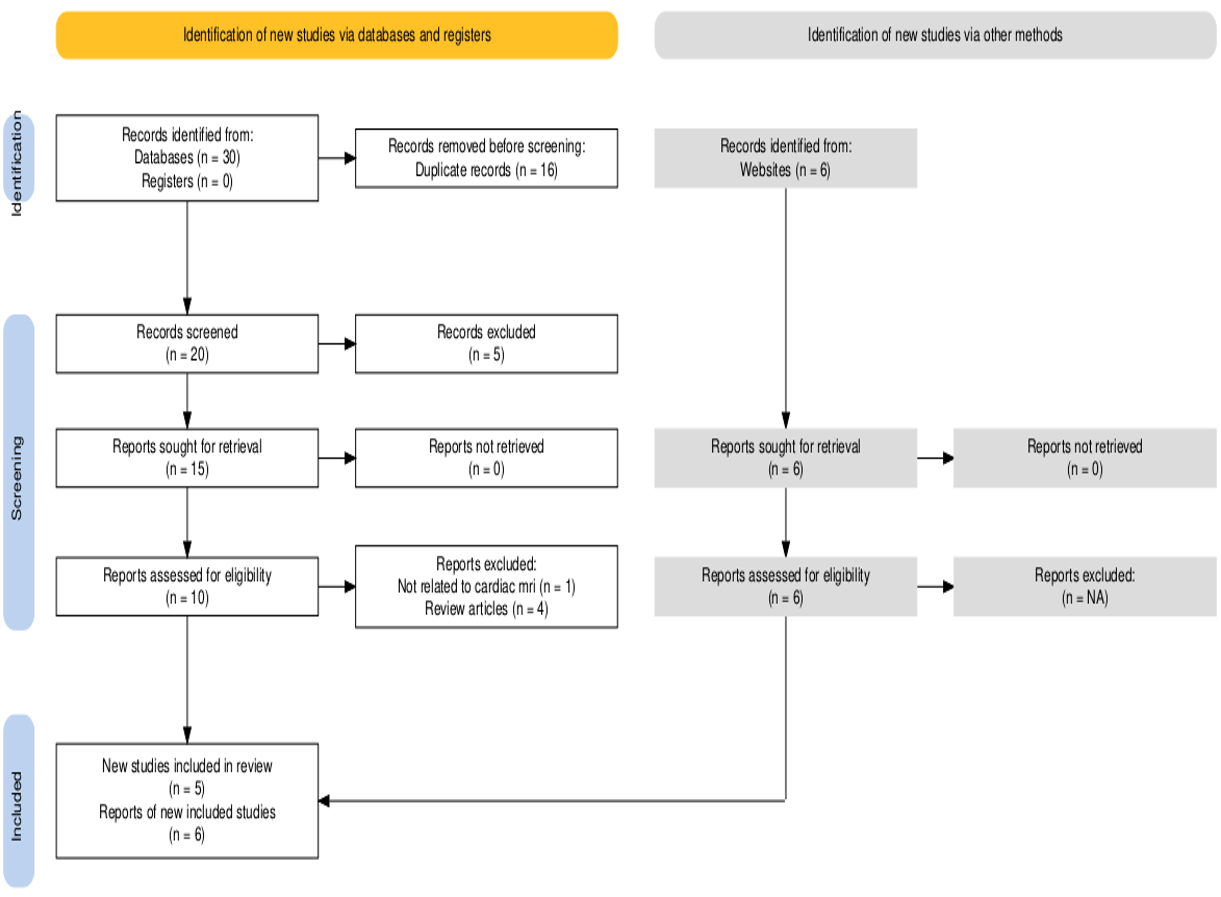
Figure 1: Prisma Flow chart
FIGURE 2
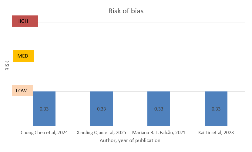
Graph 1: Risk of Bias of 4 research articles
Tables at a glance
Figures at a glance