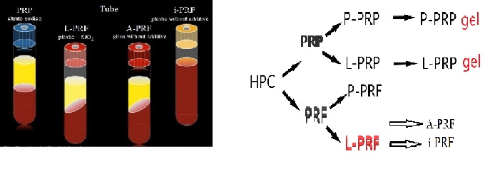
Figure 1 Platelet concentrates (HPC)
Figure 1 Platelet concentrates (HPC)
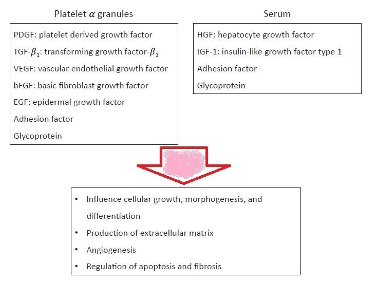
Figure 2 Function of the platelets in wound healing
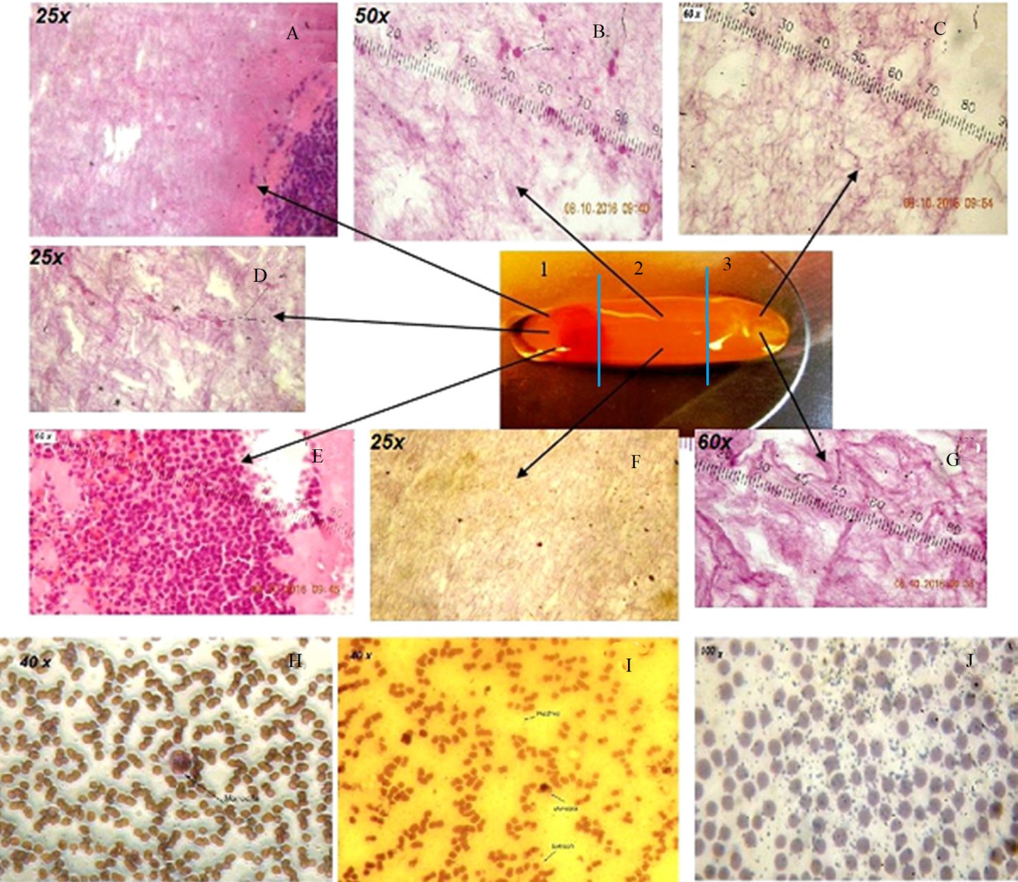
Figure 3 Horse L-PRF membrane at 0 minutes from compression (Eosin-Hematoxylin color). The L-PRF layers were fixed in 10% formalin buffered neutral solution at pH 7.2 for 48 hours and incorporated in paraffin according to the standard procedure. Twenty serial sections (7 μm thickness) of each sample were cut using a microtome. A) III proximal ingr. 25x White Blood Cells- Fibrin Reticulum; B) III average ingr. 60x Erythrocytes- Fibrin pattern; C) III distal ingr. 60x Fibrin Reticulum; D) III proximal ingr. 25x Erythrocytes-Fibrin; E) III proximal ingr. 60x Fibrin on the right, Lymphocytes in the center, Erythrocytes and neutrophil granulocytes on the left; F) III medium ingr. 25x fibrin lattice; G) III distal ingr. 60x Fibrin Reticulum; H) Red clot smear ingr. 40x presence of monocita in a carpet of erythrocytes; I) smear red clot ingr. 40x presence of erythrocytes, monocytes and platelets; J) smear red clot ingr. 100x platelets in a carpet of erythrocytes (May-Grunwald- Giemsa stain). (Crisci et al. 2017) [4].
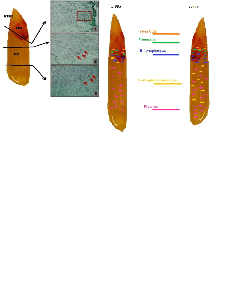
Figure 4 A-PRF (Advanced-PRF) Total scan of a fibrin clot along its longitudinal axis (Masson-Goldner staining).
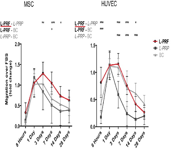
Figure 5MSC and HUVEC migration is shown in response to factors released by L-PRF, L-PRP and blood clot (BC).