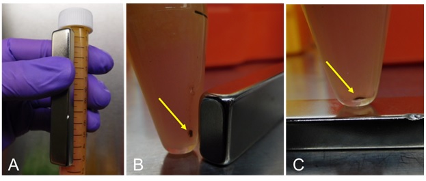
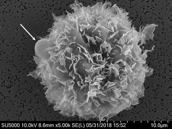
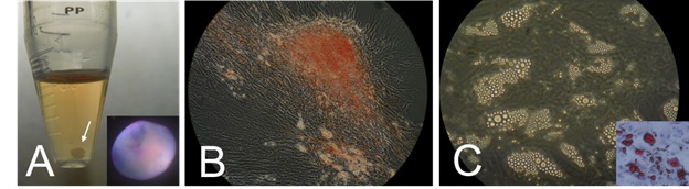
Figure 1:Paramagnetic immunobead.

Primary and secondary IgG antibodies, diagnostic of ASCs, are built onto a paramagnetic microbead, creating a paramagnetic immunobead. Layering the antibodies respectively may reduce steric hindrance and improve binding strength to conjugated ASCs.

Figure 2:Paramagnetic immunoprecipitation of ascs.

After placement of the paramagnetic immunobeads (PIBs) into the lipoaspirate, the tube containing the lipoaspirate-PIB mixture is manually held and rotated for 10 minutes. Over the next 10 minutes, a neodymium bar magnet is moved from alongside the tube to the bottom of the tube (panels A-C) to precipitate the ASC-PIBs conjugates (arrows). The lipoaspirate is then simply discarded.

Figure 3:Scanning electron microscopy.

Scanning electron micrograph of a cell phenotypically consistent with an adipose-derived stem cell conjugated to a paramagnetic immunobead (arrow).

Figure 4:Immunophenotyping of primary adipose-derived cells (ascs) isolated with paramagnetic immunobeads (pibs).

Row A: Scatter plots of cultured ASCs displaying gating to singlets.
Row B: Scatter plot and histogram of cultured ASCs displaying positive CD105 (PerCP-CyTM5.5-CD105) as part of the "Cocktail."
Row C: Scatter plot of freshly isolated ASC-PIB conjugates (aPIBs) with gating to singlets.
Row D: Scatter plot and histogram displaying positive CD105 (PerCP-CyTM5.5-CD105) as part of the "Cocktail." While FITC-CD90 also displayed similar positivity, APC-CD73 and PE-CD44 were not clearly appreciated due to the auto-fluorescence of the PIBs.

Figure 5:Culture morphology.

Bright-field micrograph of flat plate culture morphology of adipose-derived stem cells precipitated from lipoaspirate with paramagnetic immunobeads (day 14). Well-organized spheres have developed as well as large colonies. Most of the paramagnetic immunobeads have become spontaneously unconjugated. (10x, measurement bar is 250 µm).

Figure 6:Tri-lineage differentiation.

Tri-lineage differentiation of adipose-derived stem cells precipitated with paramagnetic immunobeads. Panel A: CHONDROBLAST DIFFERENTIATION: Gross chondrosphere (at white arrow) with representative indirect micrograph illustrating a sulfated proteoglycan sphere (inset) stained with Alcian Blue & Nuclear Fast Red, consistent for chondroblasts. Sphere diameter about 3mm. Panel B: OSTEOBLAST DIFFERENTIATION:Phase contrast 20x micrograph illustrating alkaline phosphatase staining with Alizarin Red, consistent for osteoblasts. Panel C: ADIPOCYTE DIFFERENTIATION: Phase contrast 10x micrograph illustrating large lipid filled droplets, consistent for adipocytes (higher power inset with lipid droplet staining by Oil-Red-O).