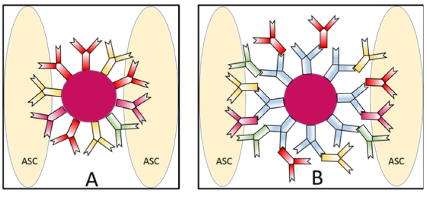
Figure 1:Paramagnetic immunobeads (PIBs).
Figure 1 A. Single core PIB structure. The paramagnetic bead (magenta center) is linked to four different antibodies (CD44, CD73, CD90, and CD105) which are diagnostic for adipose-derived stem cells (ASCs). The antibodies are attached to the bead via a Protein G linker.
Figure 1 B. Dual core PIB structure. The proteomic structure remains the same as the single core PIB except that secondary antibodies (blue) are linked initially to the PIB, and then to the identical primary antibodies used in the single core design.
Figure 2:Adipose-derived stem cell(ASC) spheroids self-assemble after ASC-paramagnetic bead (aPIB) immunoprecipitation as seen under phase-contrast microscopy at culture day 5.
A. ASC micro-spheroids (arrow).These micro-spheroids self-assembled with the aid of single core PIBs. The average micro-spheroid diameter is approximately 20 μm (+/- 5μm). The measurement bar is 25 μm (40x mag.).
B. ASC macro-spheroid (arrow). These macro-spheroids self-assembled with the aid of dual core PIBs. The average macro-spheroid diameter is approximately 220 μm (+/-25 μm). The measurement bar is 250 μm (10x mag.). [Note that the free immunobeads appear green whilst the spheroids appear dark brown (which is closer to the dark magenta color of these immunobeads)].
Figure 3:Quantities of adipose-derived stem cells (ASCs) and subsequent spheroid diameter using single core paramagnetic immunobeads (scPIBs, top panel) and dual-core paramagnetic immunobeads (dcPIBs, bottom panel)].