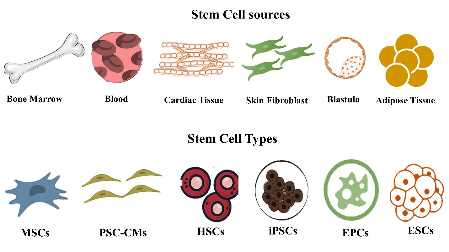
Figure1: Schematic illustration of different possible stem cell sources that are used in cardiac tissue regeneration and different stem cell types used in cardiac regeneration
Figure1: Schematic illustration of different possible stem cell sources that are used in cardiac tissue regeneration and different stem cell types used in cardiac regeneration
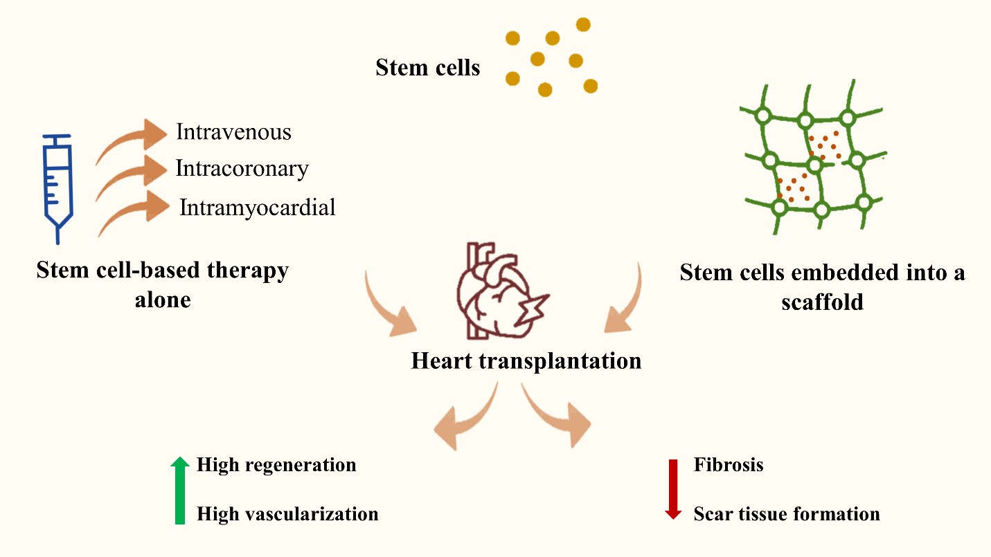
Figure2: Schematic illustration of the stem cells used as therapy in cardiac regeneration with co-administration of other materials that help in adhesion, retention, and proliferation. The stem cells can be injected intravenously, or directly into the affected cardiac tissue via intramyocardial administration. Also, it can be applied intracoronary. They can be applied alone or seeded in a scaffold. After administration, stem cells exhibit better regeneration and angiogenesis with less fibrosis and scar tissue formation
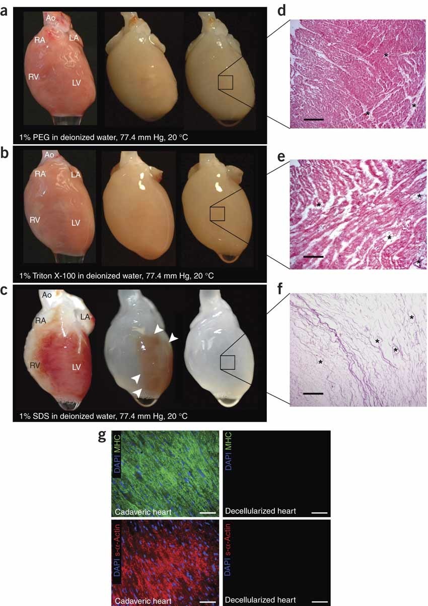
Figure3: A microscopic and macroscopic illustration of the process of rat heart decellularization using different techniques. Adapted from Ref. [27] with copyrights
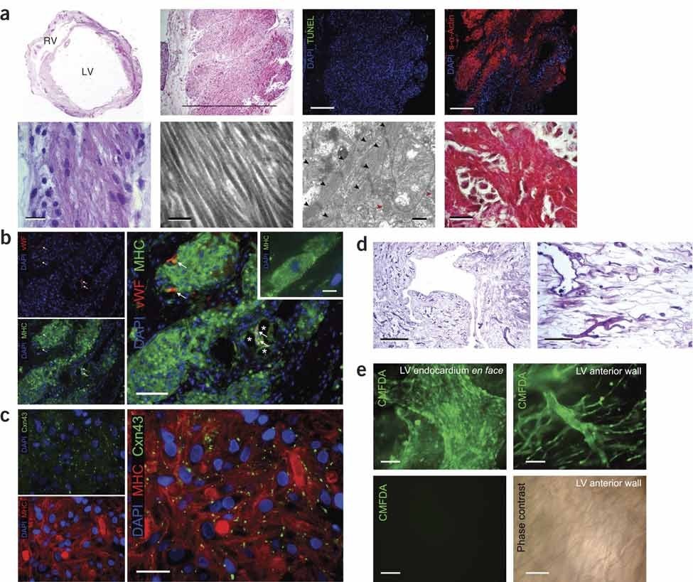
Figure4: Histological illustration of rat cardiac recellularization indicating the thickness in the ventricular wall, and mature regions of cardiomyocytes.The expression of gap-junction associated protein. Re-reendothelialization is observed, the appearance of small and large vessels. Adapted from Ref. [27] with copyrights
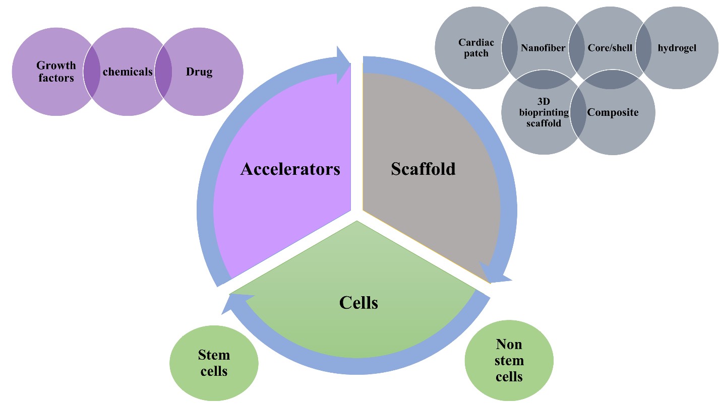
Figure5: Schematic Illustration of the cardiac tissue regeneration approaches and applications with the three major elements required for tissue engineering. 1) The scaffolds, are composed of biomaterials that are mostly natural biodegradable polymers designed by different fabrication techniques. 2) The cellular elements which mainly stem cells, can be obtained from non-stem cell sources as well and 3) biomolecules as growth factors or drugs or chemical compounds that help in accelerating the regeneration
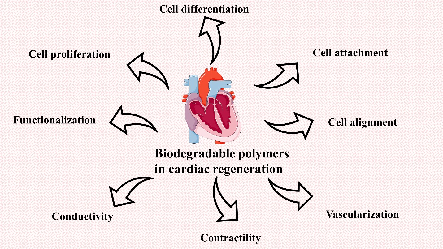
Figure6: Illustration of the main features the scaffold and polymers should possess in order to achieve appropriate and correct cardiac tissue regeneration with maintaining cardiac tissue characteristics, function, and structure
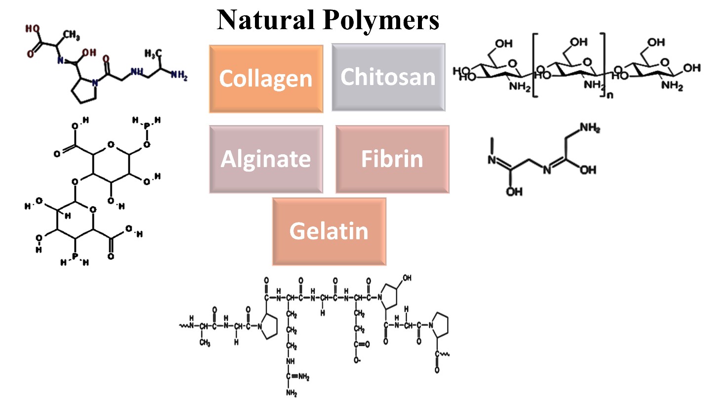
Figure7: Schematic illustration of natural biodegradable polymers that are widely used in cardiac tissue engineering
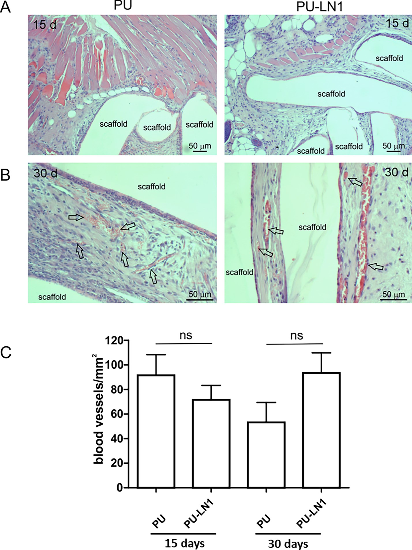
Figure8: Histological examination by hematoxylin and eosin staining of the subcutaneously implanted scaffolds in mice. The PUR-LN1 did not induce an inflammation response. Adapted from Ref [65] with copyrights
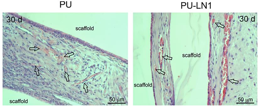
Figure9: Illustration of the blood vessels formed after scaffold transplantation in mice when inject subcutaneously and examined histologically by a light microscope. Adapted from Ref [65] with copyrights
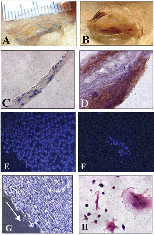
Figure10: A figure illustrates the comparison between endothelial seeded A and C and the unseeded B and D polyurethane leaflet. The seeded valve did not induce thrombosis(A), the unseeded induced abnormal gross thrombi (B). in (C) and (D) immunostaining for platelets markers CD41/CD61, the seeded showed no immunoreaction response (C) unlike the unseeded (D). Adapted from Ref [68] with copyrights
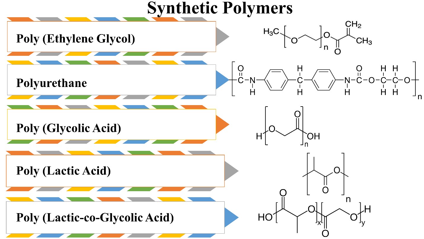
Figure11: Synthetic biodegradable polymers used in scaffold fabrication for cardiac tissue regeneration
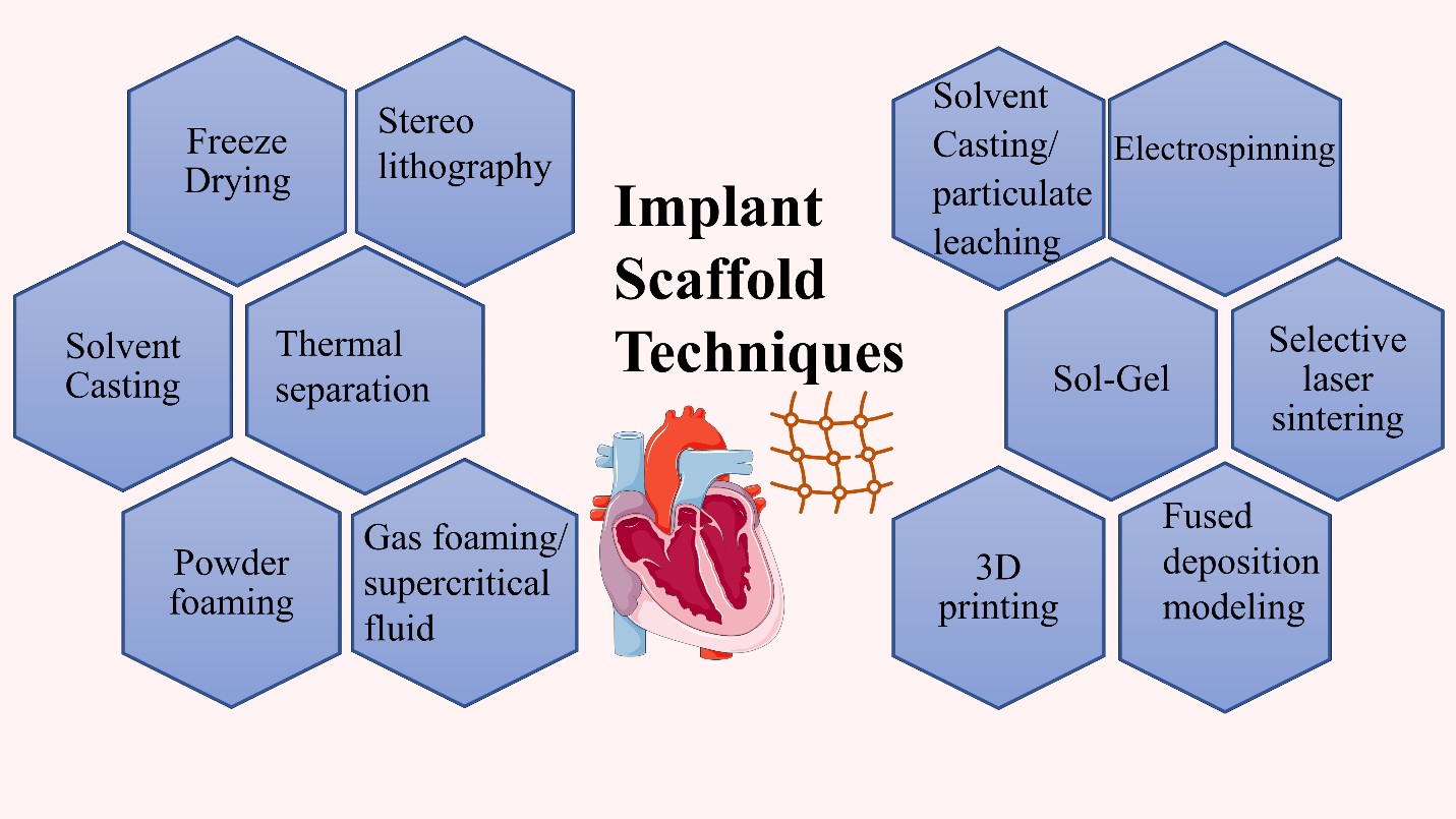
Figure12: Classification of different implant fabrication techniques used in cardiac tissue regeneration
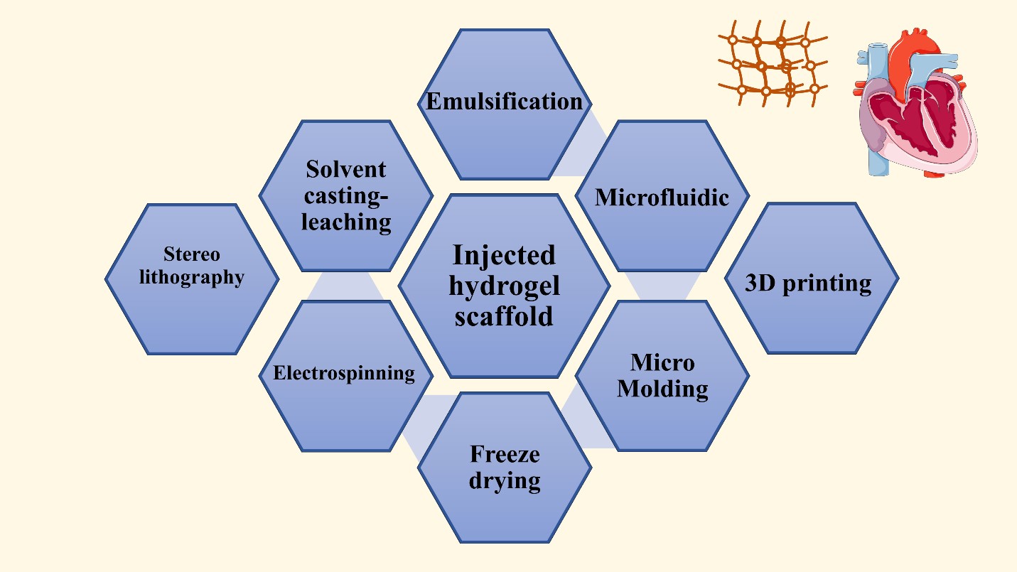
Figure13: Classification of different fabrication techniques used in cardiac tissue regeneration to obtain injected hydrogel scaffolds
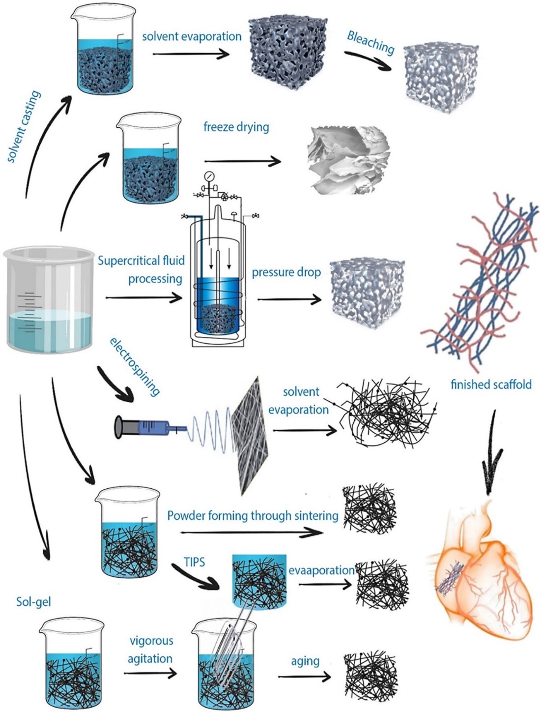
Figure14: Schematic illustration of the conventional fabrication techniques and the manufacturing methods to produce implanted scaffolds. Adapted from Ref [70] with copyrights
Figures at a glance