Cell types |
Suppliers (batch) |
Culture medium |
Human hair follicle dermal papilla cells (HHFDPC)1 |
PromoCell (403Z014.6) |
Mesenchymal stem cell medium (MSCM) +5% FBS and growth factors |
Keratinocytes of outer root sheath (KORS) |
ScienCell (9265) |
Mesenchymal stem cell medium (MSCM) +5% FBS and growth factors |
Human dermal microvascular endothelial cells (HDMEC) |
ScienCell (2622) |
Endothelial cell medium (ECM) +5% FBS and growth factors |
Human hair follicle stem cells (HFSCs) |
CliniSiences (36007-08) |
Hair follicle stem cell medium (HFSCM) with growth factors |
Table 1: Cell types
1For all experiments, HHFDPCs have been used as spheroids.
Genes |
Forward primers |
Reverse primers |
BMP2 |
5’- TTTCAATGGACGTGTCCCCG-3’ |
5’- AGCAGCAACGCTAGAAGACA-3’ |
BMP4 |
5’- GGGATTCCCGTCCAAGCTAT-3’ |
5’- ATGGCACTACGGAATGGCTC-3’ |
BMP6 |
5’- CTCAACCGCAAGAGCCTTCTG-3’ |
5’- CTTTGTGGTGTCGCTGACGA-3’ |
CD200 |
5’- GCCTGCCTCACCGTCTATG-3’ |
5’- GTGGTCCCATTTGGGTGAGA-3’ |
CD34 |
5’- TGTCTACTGCTGGTCTTGGC-3’ |
5’- AGGTGACCAGTGCAATCAGG-3’ |
cK15 |
5’- ATGAAGGAGTTCAGCAGCCA-3’ |
5’- TGGAGGCCACCTCTTTGTTC-3’ |
GPC1 |
5'-TGCCCTGACTATTGCCGAA-3' |
5'-CATGGAGTCCAGGAGGTTCCT-3' |
GPC3 |
5'-GCCCATTCTCAACAACGCCA-3' |
5'-TGTAGCCAGGCAAAGCACTA-3' |
GPC4 |
5'-AGCGGTTGCGGGAGATGTCGT-3' |
5'-AGTCACGAGACCCCGGCAGTG-3' |
GPC5 |
5'-GGCATGGTTGAACAAGTCAG-3' |
5'-GCCAGTGTCTGTTTGATGGA-3' |
GPC6 |
5'-AGAGCGACTGGAGGGGCCATT-3' |
5'-TTCAGGAGCTGAGCGGGCAGA-3' |
ITGA6 |
5’- GGACATTCTCATGCGAGCCT-3’ |
5’- ATCCACCAAGGTACTCCCGA-3’ |
ITGB1 |
5’- CGCCGCGCGGAAAAGATG-3’ |
5’- AAACACCAGCAGCCGTGTAA-3’ |
LGR5 |
5’- AACAGAAATCCCCGTCCAGG-3’ |
5’- TGTGGAGCCCATCAAAGCAT-3’ |
GAPDH |
5'-GCAGACAAGGTCCCAAAGAC-3' |
5'-ACCACCCTGACACATAAACC-3' |
TBP |
5'-TGCACAGGAGCCAAGAGTGAA-3' |
5'-CACATCACAGCTCCCCACCA-3' |
Wnt3a |
5’- CGAGTTTGGTGGGATGGTGT-3’ |
5’- AGCATGTCTTCACCTCGCAG-3’ |
Wnt5a |
5’- CTTAACCCGGTCGCTCCG-3’ |
5’- CCATCCCCAAAGCAACTCCT-3’ |
Table 2: Primers used for real-time RT-PCR
Recognized proteins |
Host and isotype |
Dilution or Concentration |
References |
Actin |
Rabbit polyclonal |
1/2000 |
A2066, Sigma-Aldrich |
BMP2 |
Rabbit polyclonal |
1/1000 |
ab14933, Abcam, Cambridge,UK |
BMP4 |
Rabbit polyclonal |
1 µg/mL |
ab39973, Abcam |
BMP6 |
Rabbit monoclonal |
1/1000 |
ab155963, Abcam |
BMPR2 |
Mouse monoclonal |
1/1000 |
ab78422, Abcam |
CD200 |
Mouse polyclonal |
1/500 |
H00004345-B01P, Bio-Techne, |
CD34 |
Rabbit polyclonal |
1/1000 |
NBP2-38321, Bio-Techne |
Fz7 |
Rabbit polyclonal |
1/1000 |
16974-1-AP, Proteintech, Rosemont, IL, USA |
GPC1 |
Rabbit polyclonal |
1/1000 |
16700-1-AP, Proteintech |
GPC4 |
Rabbit polyclonal |
1/1000 |
13048-1-AP, Proteintech |
GPC6 |
Goat polyclonal |
0.1 µg/mL |
AF2845, Bio-Techne |
LGR5 |
Rabbit polyclonal |
1 µg/mL |
NBP1-28904, Bio-Techne |
Patched 1 |
Rat monoclonal |
1 µg/mL |
MAB41051, Bio-Techne |
Shh |
Rabbit monoclonal |
1/1000 |
ab53281, Abcam |
Wnt3a |
Rabbit polyclonal |
1 µg/mL |
ab28472, Abcam |
Wnt5a |
Rabbit monoclonal |
1/1000 |
ab179824, Abcam |
Table 3: List of primary antibodies used in immunoblotting and co-immunoprecipitation experiments
The appropriate peroxidase-coupled secondary antibodies (1/10000) were the anti-rabbit NA934V (GE Healthcare Life Sciences, Marlborough, MA, USA), the anti-mouse NA931V (GE Healthcare Life Sciences), the anti-rat NA935V (GE Healthcare
Life Sciences) and the anti-goat A9452 (Sigma-Aldrich, Saint Louis, MO, USA).
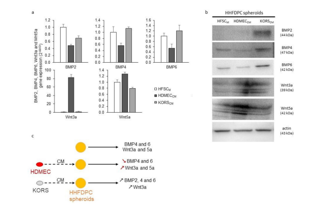
Figure 1: Effect of KORSCM and HDMECCM on growth factor expression by HHFDPCs. (a-b) Gene (a) and protein (b) expressions of BMP2, BMP4, BMP6, Wnt3a and Wnt5a in HHFDPCs.These cells were incubated in KORSCM or HDMECCM for 48 h before the analysis. N=1 experiment, n=3 replicates. (c) A summary diagram is presented to illustrate the effect of the conditioned media on the expression of BMP2, 4 and 6 as well as Wnt3a and 5a by HHFDPCs.
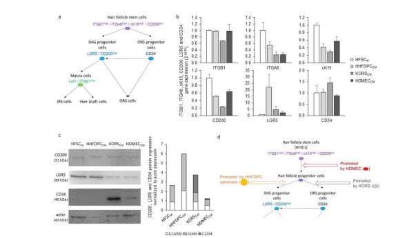
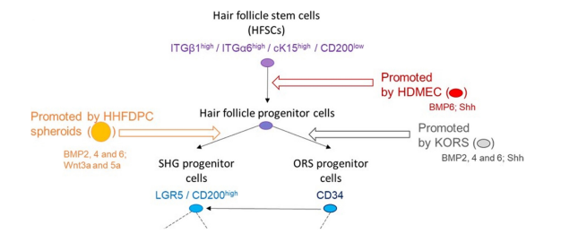
Figure 2: Effect of HHFDPCCM, KORSCM and HDMECCM on HFSC diffrentiation (a) A scheme of HFSC diffrentiation, as described in the literature, is illustrated. (b) ITGB1, ITGA6, cK15, CD200, LGR5 and CD34 gene expression in HFSCs was analyzed by RT-qPCR. N=2 independent experiments, n=3 replicates per experiment. (c) Protein expression and quantification of CD200, LGR5 and CD34 in the HFSCs was determined by Western immunoblotting.These HFSCs were incubated in HHFDPCCM, KORSCM or HDMECCM for 48 h before the analysis. (d) A summary diagram is presented to illustrate the effect of the conditioned media on HFSCs diffrentiation.
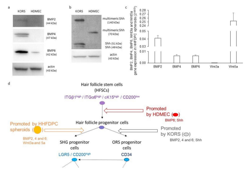
Figure 3: KORS, HDMECs and HHFDPCs express growth factors known to be involved in the regulation of HFSC behavior. (a-b) Protein expression of BMP2, BMP4, BMP6 (a) and Shh (b) in the HFSCs was determined by Western immunoblotting. (c) BMP2, BMP4, BMP6, Wnt3a and Wnt5a gene expression in HHFDPC spheroids were analyzed by RT-qPCR. N=2 independent experiments, n=3 replicates per experiment. (d) A summary diagram is presented to illustrate the effect of the conditioned media on HFSCs, and the growth factors expressed by KORS, HDMECs and HHFDPCs.
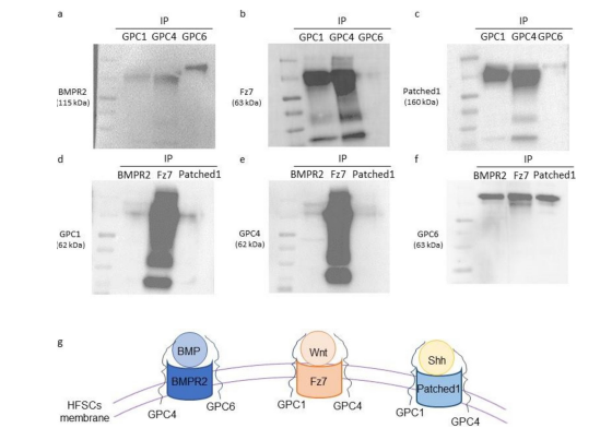
Figure 4: GPC1, GPC4 and GPC6 interact with BMPR2, Fz7 and Patched 1. (a-c) A whole cell protein extract (25 μg) from the HFSCs was precipitated using anti-GPC1, anti-GPC4 or anti-GPC6 antibody. Anti-BMPR2 (a), anti-Fz7 (b) or anti-Patched 1 (c) antibodies were used to reveal the membrane. (d-f) A reverse immunoprecipitation assay, corresponding to the co-immunoprecipitation shown in Figure 4a-c, was conducted from HFSC whole cell protein extracts using anti-BMPR2, anti-Fz7 or anti-- Patched 1 antibody. Then, the isolated immunocomplexes were immunoblotted using anti-GPC1 (d), anti-GPC4 (e) or anti-GPC6 (f) antibodies. (g) A summary diagram is presented to illustrate the interaction of GPC1, 4 and 6 with BMPR2, Fz7 and Patched 1.
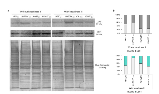
Figure 5: Effect of heparinase III incubation on HFSC diffrentiation induced by HHDCFCM, KORSCM or HDMECCM. Protein expression and quantification of LGR5 and CD34 in the HFSCs was determined by Western immunoblotting.The cells were incubated in HHFDPCCM, KORSCM or HDMECCM without or with heparinase III treatment for 48 h before the analysis.
Tables at a glance
Figures at a glance