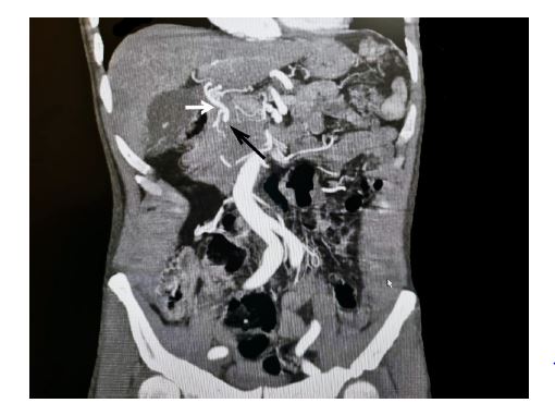
Figure 1: Black Arrow: Coronal view of the GDA aneurysm with scant amount of hematoma around it in the pancreatic head region, White Arrow: The Proper Hepatic Artery
Figure 1: Black Arrow: Coronal view of the GDA aneurysm with scant amount of hematoma around it in the pancreatic head region, White Arrow: The Proper Hepatic Artery
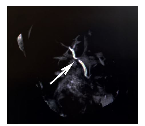
Figure 2: The MRCP shows the indentation of the aneurysm on the biliary system (Common Bile Duct)
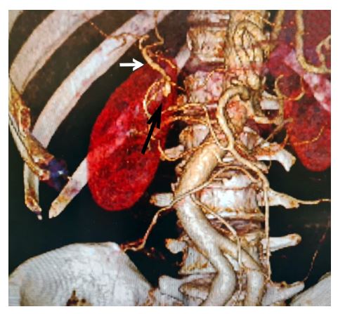
Figure 3: Three-dimensional CT angiography of GDA's aneurysmal site. Black Arrow indicated the sac, note the variation of the common hepatic artery originating from SMA, No inflow from celiac trunk. White arrow: The Proper Hepatic Artery

Figure 4: Post operation Triphasic CT scan, after reconstruction of hepatic artery, Black Arrow: End to side arterial anastomosis of proper hepatic artery to splenic artery. White arrow: The Proper Hepatic Artery
Figures at a glance