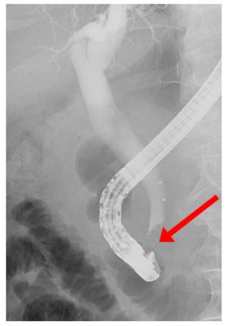
Figure 1: Endoscopic retrograde cholangioscopy
In the distal hepatocholedochal duct there seems to be a contrast media sparing concrement or adenoma (red arrow).
Figure 1: Endoscopic retrograde cholangioscopy
In the distal hepatocholedochal duct there seems to be a contrast media sparing concrement or adenoma (red arrow).
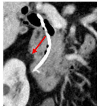
Figure 2: CT-Scan of the duodenal region
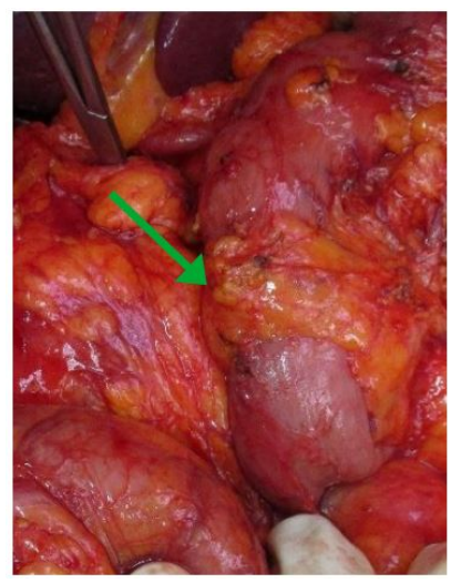
Figure 3: Intraoperative Picture of annular pancreas The descendent part of duodenum is totally encircled with pancreatic tissue (green arrow). Ampulla of vater has not been detectable.
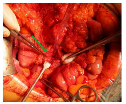
Figure 4: Photography of the ampulla of vater
In the center of the duodenotomy the ampulla has been exposed (green arrow). The adenoma is not shown.
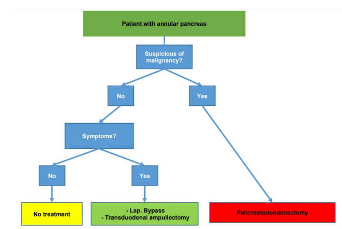
Figure 5: Treatment scheme for annular pancreas patients
Figures at a glance