The 52-year-old female patient was sent to the gastrointestinal clinic due to an abdominal ultrasonography-detected cystic pancreas lesion that was discovered in an outside hospital. The biochemical tests and complete blood count were all within normal ranges, with the exception of high levels of glucose (144 mg/dL), triglycerides (578 U/mL), and hemoglobin (7.5). An abdominal computed tomography scan revealed a cystic mass lesion measuring 5 by 4 cm, surrounded by granular calcification.
Singh S, et al [28].
A 44-year-old lady, who was obese and otherwise in good condition, appeared with a left-sided abdominal mass that she had been carrying for the previous year. She had no significant past medical history. She denied experiencing any of the following symptoms: chills, fever, jaundice, anorexia, flushing, weight loss, fullness, diarrhea, nausea, and/or vomiting. With a carcinoembryonic antigen (CEA) level of 0.9 ng/ml and a carbohydrate antigen 19-9 (CA 19-9) level of 5U/ml, the tumor markers were within normal limits. A very large mass with solid and cystic features as well as calcifications coming from the body and tail of the pancreas, which were highly deformed and irregular, were seen on computerized tomography.
Gursev Sandlas, et al [29].
An 11-year-old girl who was otherwise in good health arrived with acute abdominal pain that started suddenly and persisted for a single day. The patient revealed a history of repeated episodes of stomach pain during the previous six months upon more direct inquiry. The general checkup revealed nothing noteworthy. It was a soft abdomen. Although close by, the stomach and left kidney were unaffected. Although the mass had a clearly defined border, it was unable to identify the organ of origin.
Table 1: Summarizing key characteristics of SPN
Tables at a glance
Table 1
Figures at a glance
Figure 1Figure 2Figure 3Figure 4
FIGURE 1
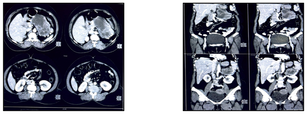
Figure 1: Computed tomography scan of the abdomen showing A large heterogeneous mass
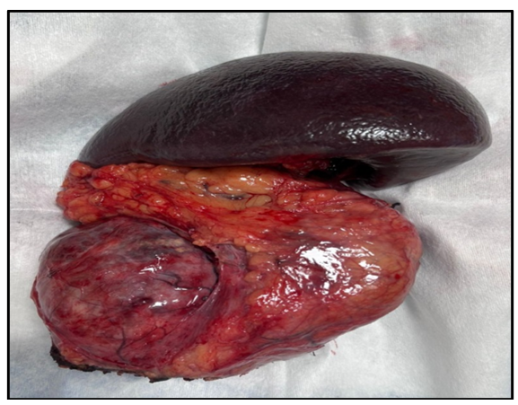
FIGURE 2
Figure 2:Post operative specimen showing tumor in the body of pancreas with spleen
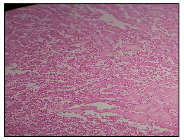
FIGURE 3
Figure 3:Tumor cells have a moderate amount of eosinophilic cytoplasm and perinuclear vacuoles
FIGURE 4
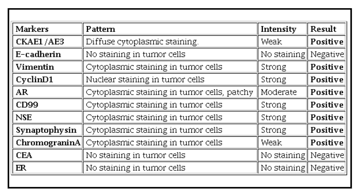
Figure 4: Interpretation of Immunohistochemistry findings
Tables at a glance
Figures at a glance