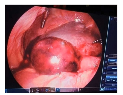
Figure 1: Intraoperative image of necrotic, dilated gallbladder
Figure 1: Intraoperative image of necrotic, dilated gallbladder
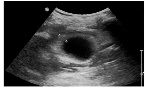
Figure 2: Right upper quadrant ultrasound demonstrates a distended gallbladder with wall thickening, sludge, and pericholecystic fluid but without calculi
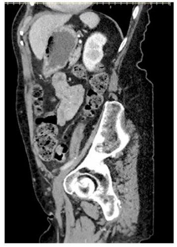
Figure 3: Sagittal CT slice demonstrates the presence of intramural air within the dilated gallbladder
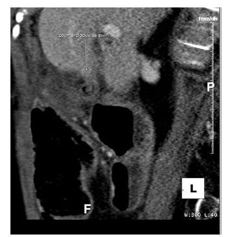
Figure 4: The presence of a "beak" and "swirl" sign at the location of the gallbladder neck
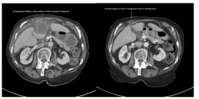
Figure 5: Axial CT slice demonstrates the relative displacement of the gallbladder compared to its location six months prior
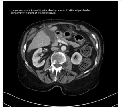
Figure 6: Axial CT scan slice that demonstrates previous normal anatomical location of gallbladder
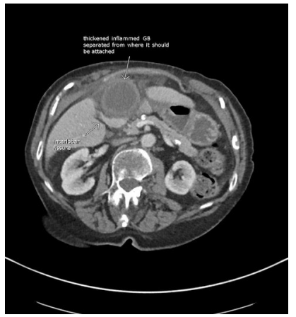
Figure 7: Axial CT slice revealing the distance of separation of inflamed gallbladder from the interlobar fissure, displacement anteriorly and medially to its normal attachment
Figures at a glance