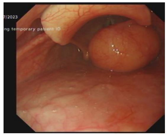
Figure 1A: OGD demonstrating right supraglottic mass obscuring laryngeal inlet and vocal cords. Only the anterior commissure of the true vocal cords can be visualized
Figure 1A: OGD demonstrating right supraglottic mass obscuring laryngeal inlet and vocal cords. Only the anterior commissure of the true vocal cords can be visualized
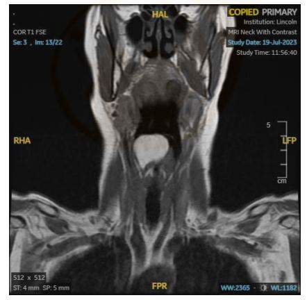
Figure 1B: MRI Neck with Contrast T1 weighted Coronal View showing a hyperintense lesion 266 situated in the right supraglottic region and crossing the midline of the laryngeal inlet
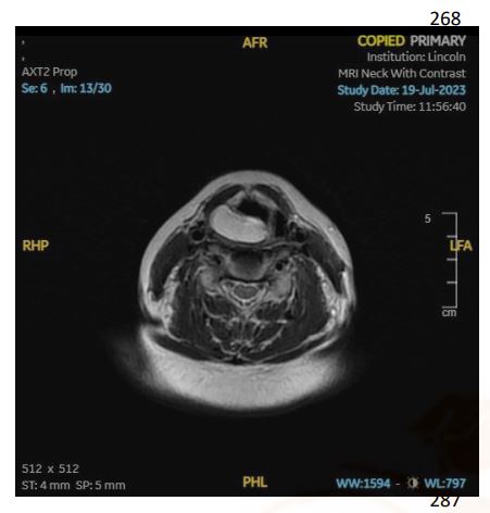
Figure 1C: MRI Neck with Contrast T1 weighted Axial View showing a hyperintense lesion situated at the level of the right supraglottis
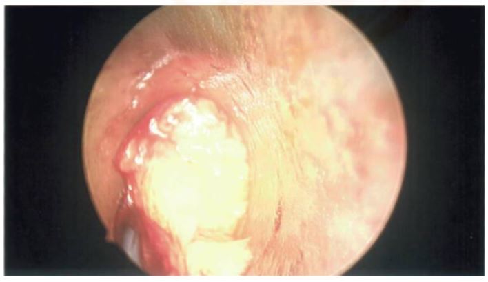
Figure 2A: Laryngoscopy showing a lipomatous lesion within the right supraglottis
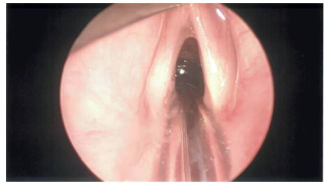
Figure 2B: Laryngoscopy demonstrating normal true and false vocal cords with endotracheal tube in situ
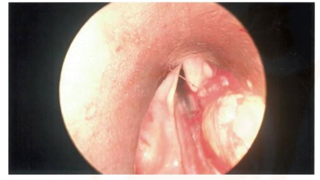
Figure 2C: Laryngoscopy showing the lipomatous lesion in the right supraglottic region encroaching on the endotracheal tube. This was just before excision of the lesion
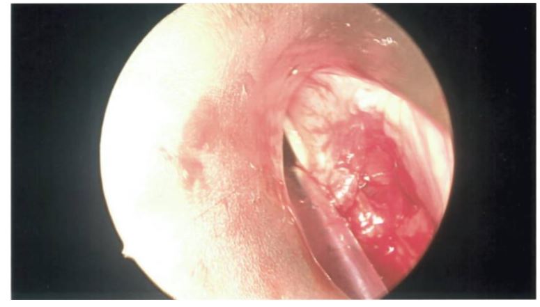
Figure 2D: Laryngoscopy demonstrating the right supraglottic region post removal of the lipomatous lesion
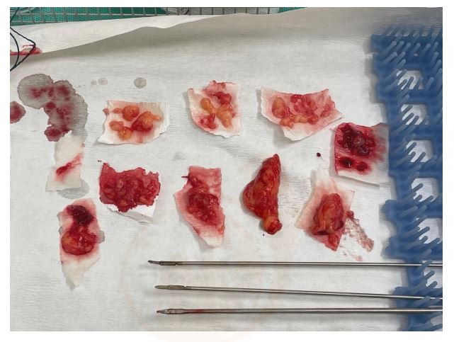
Figure 3: Photograph showing constituent parts of the lipomatous lesion excised
Figures at a glance