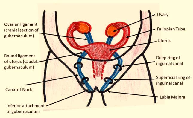
Figure 1: Diagrammatic depiction of embryological attachment of gubernaculum that occurs from female gynecological organs to labia majora.
Figure 1: Diagrammatic depiction of embryological attachment of gubernaculum that occurs from female gynecological organs to labia majora.
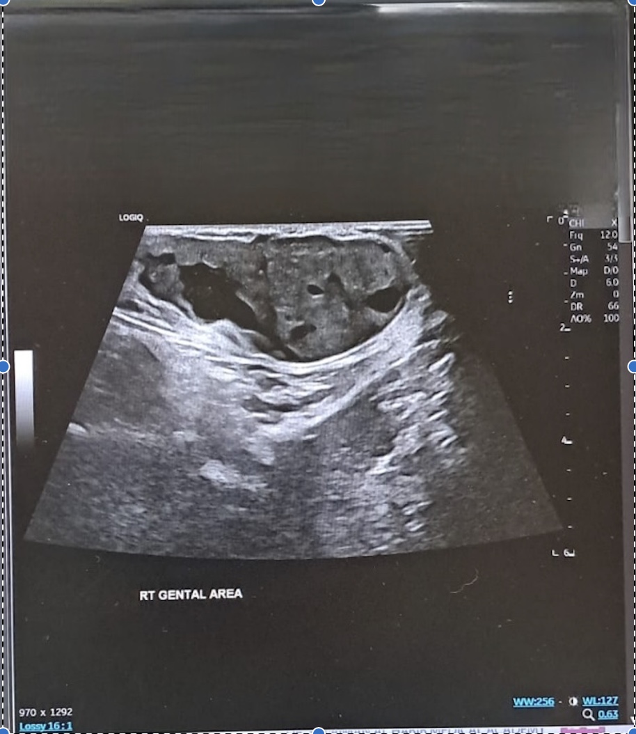
Figure 2: The ultrasound shows the hydrocele of Nuck's canal. The arrow illustrates a comma-shaped hydrocele attenuated by the fluid.
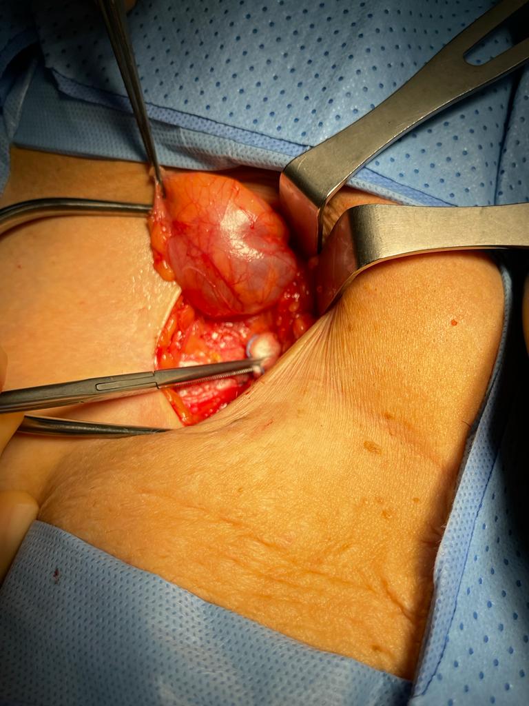
Figure 3: Intraoperative picture of a hydrocele of the canal of Nuck
Tables at a glance
Figures at a glance