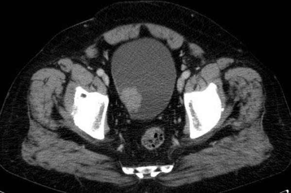
Figure1: Transverse CT-scan image showing a 3cm mass in the posterior urinary bladder wall
Figure1: Transverse CT-scan image showing a 3cm mass in the posterior urinary bladder wall
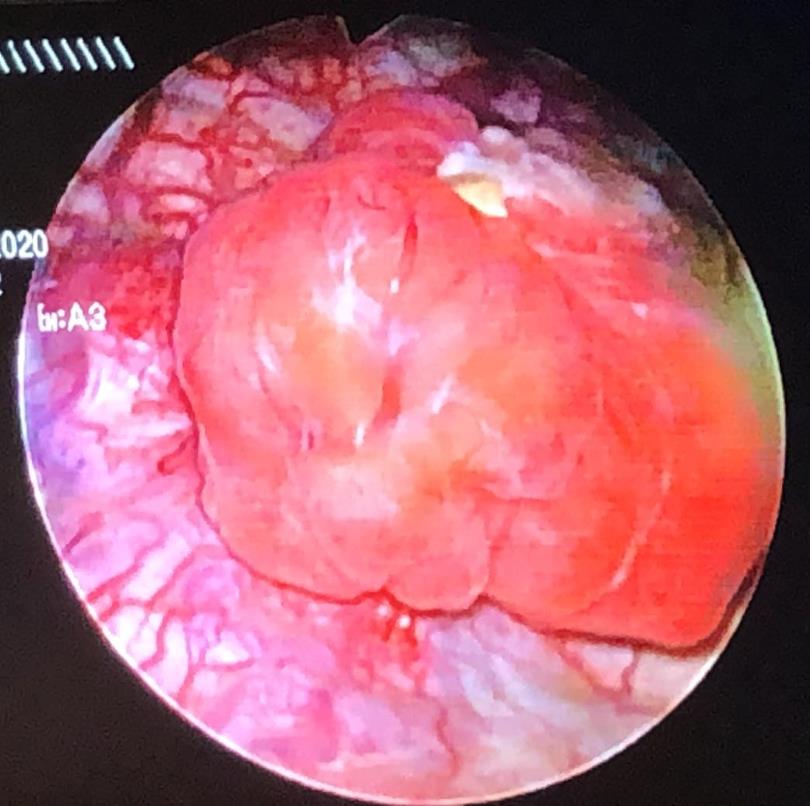
Figure2: Endoscopic image of PUB showing a mass covered by smooth vesical mucosa
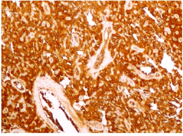
Figure3: Tumor cells are strongly stained with synaptophysin (×200)
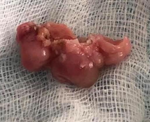
Figure4: Macroscopic view of the resected bladder paraganglioma. the tumor is red-brown in color, lobulated and well-circumscribed
Figures at a glance