Figure 1: 2D ultrasound image of the perineal mass (transvaginal)
Figure 1: 2D ultrasound image of the perineal mass (transvaginal)
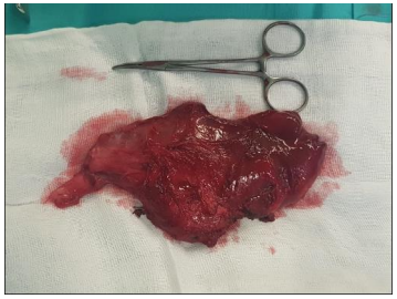
Figure 2: 2D ultrasound image of the perineal mass in close contact with the external anal sphincter (introital)
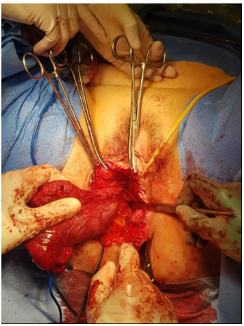
Figure 3: 3D ultrasound of the perineal mass displacing the puborrectal component of the levator ani muscle (introital)
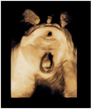
Figure 4: Surgery to remove the perineal mass
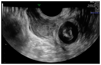
Figure 5: The surgical specimen after its removal
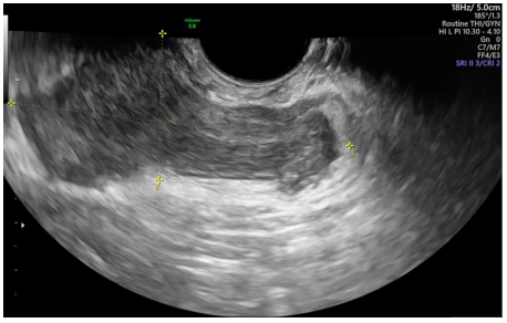
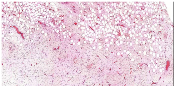
Figure 6: Microscopic section of the tumour showing peripheral adipose tissue
Figures at a glance