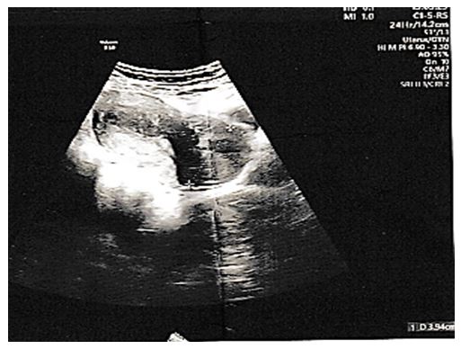
Figure 1: Ultrasound image objectifying the cervical mass
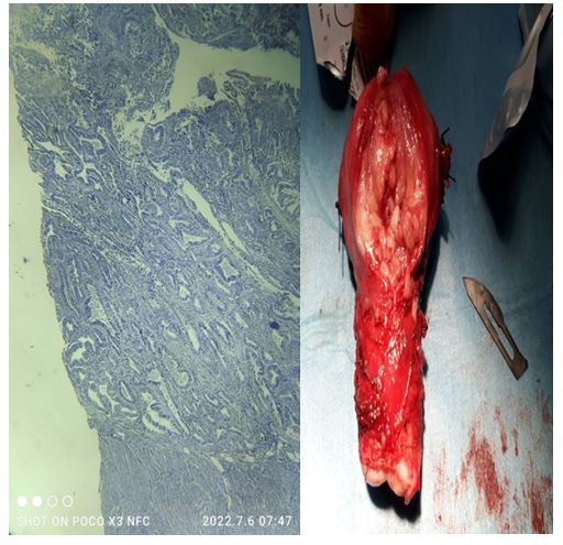
Figure 2: Left: an authentic photo of the operating room. Right: an electron microscope photo of the operating room
Figures at a glance