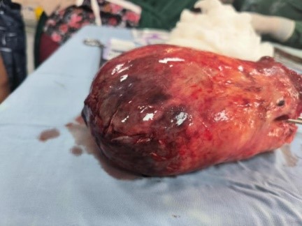
Figure 1: Gross specimen of the submucosal uterine leiomyoma after abdominal myomectomy, showing a large, congested and partially necrotic surface
Figure 1: Gross specimen of the submucosal uterine leiomyoma after abdominal myomectomy, showing a large, congested and partially necrotic surface
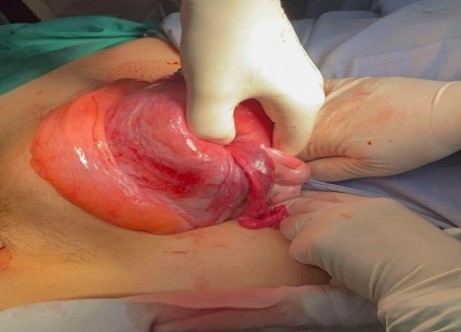
Figure 2: Intraoperative view through the suprapubic laparotomy demonstrating the inverted uterine fundus being exteriorized and manipulated prior to correction of the uterine inversion
Figures at a glance